 Movie
Movie Controller

[English] 日本語
 Yorodumi
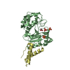
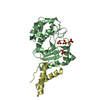
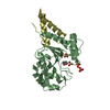
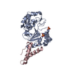
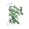
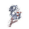
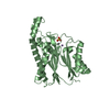
- PDB-4oda: Crystal structure of the vaccinia virus DNA polymerase holoenzyme... -
+ Open data

- Basic information
| Entry | Database: PDB / ID: 4oda | ||||||
|---|---|---|---|---|---|---|---|
| Title | Crystal structure of the vaccinia virus DNA polymerase holoenzyme subunit D4 in complex with the A20 N-terminus | ||||||
 Components Components |
| ||||||
 Keywords Keywords | HYDROLASE/REPLICATION / DNA polymerase processivity factor /  DNA / DNA polymerase E9 / HYDROLASE-REPLICATION complex DNA / DNA polymerase E9 / HYDROLASE-REPLICATION complex | ||||||
| Function / homology |  Function and homology informationuracil-DNA glycosylase / viral DNA genome replication / uracil DNA N-glycosylase activity / DNA replication / DNA repair / DNA binding Function and homology informationuracil-DNA glycosylase / viral DNA genome replication / uracil DNA N-glycosylase activity / DNA replication / DNA repair / DNA bindingSimilarity search - Function | ||||||
| Biological species |   Vaccinia virus Vaccinia virus | ||||||
| Method | X-RAY DIFFRACTION / SYNCHROTRON / MOLECULAR REPLACEMENT / Resolution: 2.2 Å | ||||||
 Authors Authors | Contesto-Richefeu, C. / Tarbouriech, N. / Brazzolotto, X. / Burmeister, W.P. / Iseni, F. | ||||||
 Citation Citation | Journal: Plos Pathog. / Year: 2014 Title: Crystal structure of the vaccinia virus DNA polymerase holoenzyme subunit d4 in complex with the a20 N-terminal domain. Authors: Contesto-Richefeu, C. / Tarbouriech, N. / Brazzolotto, X. / Betzi, S. / Morelli, X. / Burmeister, W.P. / Iseni, F. | ||||||
| History |
|
- Structure visualization
| Structure viewer | Molecule: MolmilJmol/JSmol |
|---|
- Downloads & links
-Download
| PDBx/mmCIF format | 4oda.cif.gz | 234.2 KB | Display | PDBx/mmCIF format |
|---|---|---|---|---|
| PDB format | pdb4oda.ent.gz | 189.9 KB | Display | PDB format |
| PDBx/mmJSON format | 4oda.json.gz | Tree view | PDBx/mmJSON format | |
| Others |  Other downloads Other downloads |
-Validation report
| Arichive directory | https://data.pdbj.org/pub/pdb/validation_reports/od/4odaftp://data.pdbj.org/pub/pdb/validation_reports/od/4oda | HTTPS FTP |
|---|
-Related structure data
| Related structure data | 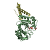 4od8SC S: Starting model for refinement C: citing same article ( |
|---|---|
| Similar structure data |


-Links
 PDBj
PDBj

- Assembly
| Deposited unit | 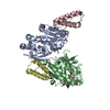
| |||||||||
|---|---|---|---|---|---|---|---|---|---|---|
| 1 | 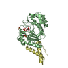
| |||||||||
| 2 | 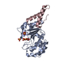
| |||||||||
| Unit cell |
| |||||||||
| Components on special symmetry positions |
|
-Components
| #1: Protein | / UDG Mass: 26729.486 Da / Num. of mol.: 2 Source method: isolated from a genetically manipulated source Source: (gene. exp.) Vaccinia virus / Strain: Copenhagen / Gene: D4R, UNG / Plasmid: pET-Duet1 / Production host:  Escherichia coli (E. coli) / Strain (production host): BL21 / References: UniProt: P20536, uracil-DNA glycosylase Escherichia coli (E. coli) / Strain (production host): BL21 / References: UniProt: P20536, uracil-DNA glycosylase#2: Protein | Mass: 5807.586 Da / Num. of mol.: 2 / Fragment: N-terminal residues 1-50 Source method: isolated from a genetically manipulated source Source: (gene. exp.) Vaccinia virus / Strain: Copenhagen / Gene: A20R / Plasmid: pET-Duet1 / Production host: Escherichia coli (E. coli) / Strain (production host): BL21 / References: UniProt: P20995#3: Chemical | ChemComp-SO4 / Sulfate  Mass: 96.063 Da / Num. of mol.: 6 / Source method: obtained synthetically / Formula: SO4 Mass: 96.063 Da / Num. of mol.: 6 / Source method: obtained synthetically / Formula: SO4#4: Chemical | Glycerol  Mass: 92.094 Da / Num. of mol.: 2 / Source method: obtained synthetically / Formula: C3H8O3 Mass: 92.094 Da / Num. of mol.: 2 / Source method: obtained synthetically / Formula: C3H8O3#5: Water | ChemComp-HOH / | Water Mass: 18.015 Da / Num. of mol.: 154 / Source method: isolated from a natural source / Formula: H2O Mass: 18.015 Da / Num. of mol.: 154 / Source method: isolated from a natural source / Formula: H2O |
|---|
-Experimental details
-Experiment
| Experiment | Method: X-RAY DIFFRACTION / Number of used crystals: 1 |
|---|
- Sample preparation
| Crystal | Density Matthews: 2.79 Å3/Da / Density % sol: 55.98 % |
|---|---|
| Crystal grow | Temperature: 293 K / Method: vapor diffusion, hanging drop / pH: 8.7 Details: 100 mM bicine, 1.5 M ammonium sulfate, pH 8.7, VAPOR DIFFUSION, HANGING DROP, temperature 293K |
-Data collection
| Diffraction | Mean temperature: 100 K |
|---|---|
| Diffraction source | Source: SYNCHROTRON / Site: ESRF  / Beamline: ID14-4 / Wavelength: 0.9394 Å / Beamline: ID14-4 / Wavelength: 0.9394 Å |
| Detector | Type: ADSC QUANTUM 315r / Detector: CCD / Date: Sep 9, 2012 / Details: Si111 monochromator and toroidal mirror |
| Radiation | Monochromator: Si 111 CHANNEL CUT / Protocol: SINGLE WAVELENGTH / Monochromatic (M) / Laue (L): M / Scattering type: x-ray |
| Radiation wavelength | Wavelength: 0.9394 Å / Relative weight: 1 |
| Reflection | Resolution: 2.2→54.02 Å / Num. all: 37537 / Num. obs: 37537 / % possible obs: 99.7 % / Redundancy: 5.3 % / Rmerge(I) obs: 0.075 / Net I/σ(I): 13.2 |
| Reflection shell | Resolution: 2.2→2.32 Å / Redundancy: 5.3 % / Rmerge(I) obs: 0.516 / Mean I/σ(I) obs: 4 / % possible all: 99.9 |
- Processing
| Software |
| ||||||||||||||||||||||||||||||||||||||||||||||||||||||||||||||||||||||||||||||||||||||||||||||||||||||||||||||||||||||||||||||||||||||||||||||||||||||||||||||||||||||||||||||||||||||
|---|---|---|---|---|---|---|---|---|---|---|---|---|---|---|---|---|---|---|---|---|---|---|---|---|---|---|---|---|---|---|---|---|---|---|---|---|---|---|---|---|---|---|---|---|---|---|---|---|---|---|---|---|---|---|---|---|---|---|---|---|---|---|---|---|---|---|---|---|---|---|---|---|---|---|---|---|---|---|---|---|---|---|---|---|---|---|---|---|---|---|---|---|---|---|---|---|---|---|---|---|---|---|---|---|---|---|---|---|---|---|---|---|---|---|---|---|---|---|---|---|---|---|---|---|---|---|---|---|---|---|---|---|---|---|---|---|---|---|---|---|---|---|---|---|---|---|---|---|---|---|---|---|---|---|---|---|---|---|---|---|---|---|---|---|---|---|---|---|---|---|---|---|---|---|---|---|---|---|---|---|---|---|---|
| Refinement | Method to determine structure: MOLECULAR REPLACEMENT Starting model: 4OD8 Resolution: 2.2→44.29 Å / Cor.coef. Fo:Fc: 0.957 / Cor.coef. Fo:Fc free: 0.934 / SU B: 12.489 / SU ML: 0.154 / Cross valid method: THROUGHOUT / ESU R: 0.222 / ESU R Free: 0.196 / Stereochemistry target values: MAXIMUM LIKELIHOOD / Details: HYDROGENS HAVE BEEN ADDED IN THE RIDING POSITIONS
| ||||||||||||||||||||||||||||||||||||||||||||||||||||||||||||||||||||||||||||||||||||||||||||||||||||||||||||||||||||||||||||||||||||||||||||||||||||||||||||||||||||||||||||||||||||||
| Solvent computation | Ion probe radii: 0.8 Å / Shrinkage radii: 0.8 Å / VDW probe radii: 1.2 Å / Solvent model: MASK | ||||||||||||||||||||||||||||||||||||||||||||||||||||||||||||||||||||||||||||||||||||||||||||||||||||||||||||||||||||||||||||||||||||||||||||||||||||||||||||||||||||||||||||||||||||||
| Displacement parameters | Biso mean: 47.682 Å2
| ||||||||||||||||||||||||||||||||||||||||||||||||||||||||||||||||||||||||||||||||||||||||||||||||||||||||||||||||||||||||||||||||||||||||||||||||||||||||||||||||||||||||||||||||||||||
| Refinement step | Cycle: LAST / Resolution: 2.2→44.29 Å
| ||||||||||||||||||||||||||||||||||||||||||||||||||||||||||||||||||||||||||||||||||||||||||||||||||||||||||||||||||||||||||||||||||||||||||||||||||||||||||||||||||||||||||||||||||||||
| Refine LS restraints |
|