 Movie
Movie Controller

[English] 日本語
 Yorodumi
- PDB-1xt8: Crystal Structure of Cysteine-Binding Protein from Campylobacter ... -
+ Open data

- Basic information
| Entry | Database: PDB / ID: 1xt8 | ||||||
|---|---|---|---|---|---|---|---|
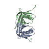
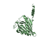
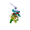
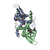
| Title | Crystal Structure of Cysteine-Binding Protein from Campylobacter jejuni at 2.0 A Resolution | ||||||
 Components Components | putative amino-acid transporter periplasmic solute-binding protein | ||||||
 Keywords Keywords |  TRANSPORT PROTEIN / ABC transport / cysteine uptake / amino-acid transporter periplasmic solute-binding protein / Campylobacter jejuni / SPINE / Structural Genomics / Structural Proteomics in Europe TRANSPORT PROTEIN / ABC transport / cysteine uptake / amino-acid transporter periplasmic solute-binding protein / Campylobacter jejuni / SPINE / Structural Genomics / Structural Proteomics in Europe | ||||||
| Function / homology |  Function and homology information Function and homology informationBacterial periplasmic substrate-binding proteins / Bacterial extracellular solute-binding proteins, family 3 / Solute-binding protein family 3/N-terminal domain of MltF / Periplasmic binding protein-like II / D-Maltodextrin-Binding Protein; domain 2 / 3-Layer(aba) Sandwich / Alpha Beta Similarity search - Domain/homology | ||||||
| Biological species |   Campylobacter jejuni (Campylobacter) Campylobacter jejuni (Campylobacter) | ||||||
| Method | X-RAY DIFFRACTION / SYNCHROTRON / MOLECULAR REPLACEMENT / Resolution: 2 Å | ||||||
 Authors Authors | Muller, A. / Thomas, G.H. / Horler, R. / Brannigan, J.A. / Blagova, E. / Levdikov, V.M. / Fogg, M.J. / Wilson, K.S. / Wilkinson, A.J. / Structural Proteomics in Europe (SPINE) | ||||||
 Citation Citation | Journal: Mol.Microbiol. / Year: 2005 Title: An ATP-binding cassette-type cysteine transporter in Campylobacter jejuni inferred from the structure of an extracytoplasmic solute receptor protein. Authors: Muller, A. / Thomas, G.H. / Horler, R. / Brannigan, J.A. / Blagova, E. / Levdikov, V.M. / Fogg, M.J. / Wilson, K.S. / Wilkinson, A.J. | ||||||
| History |
| ||||||
| Remark 999 | SEQUENCE Regarding residues -23 to 0, predicted Campylobacter jejuni leader peptide ...SEQUENCE Regarding residues -23 to 0, predicted Campylobacter jejuni leader peptide MKKILLSVLTAFVAVVLAA in the database sequence was replaced by pelB leader peptide MKYLLPTAAAGLLLLAAQPAMAMA. |
- Structure visualization
| Structure viewer | Molecule: MolmilJmol/JSmol |
|---|
- Downloads & links
-Download
| PDBx/mmCIF format | 1xt8.cif.gz | 118.2 KB | Display | PDBx/mmCIF format |
|---|---|---|---|---|
| PDB format | pdb1xt8.ent.gz | 90.3 KB | Display | PDB format |
| PDBx/mmJSON format | 1xt8.json.gz | Tree view | PDBx/mmJSON format | |
| Others |  Other downloads Other downloads |
-Validation report
| Arichive directory | https://data.pdbj.org/pub/pdb/validation_reports/xt/1xt8ftp://data.pdbj.org/pub/pdb/validation_reports/xt/1xt8 | HTTPS FTP |
|---|
-Related structure data
| Related structure data | 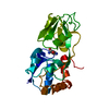 1wdnS S: Starting model for refinement |
|---|---|
| Similar structure data |


-Links
 PDBj
PDBj

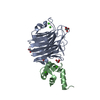
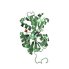
- Assembly
| Deposited unit | 
| |||||||||
|---|---|---|---|---|---|---|---|---|---|---|
| 1 | 
| |||||||||
| 2 | 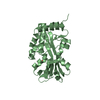
| |||||||||
| Unit cell |
| |||||||||
| Components on special symmetry positions |
|
-Components
| #1: Protein | Mass: 32449.967 Da / Num. of mol.: 2 Source method: isolated from a genetically manipulated source Source: (gene. exp.) Campylobacter jejuni (Campylobacter) / Gene: Cj0982c / Plasmid: pET22b / Production host: Escherichia coli (E. coli) / Strain (production host): BL21 Gold / References: GenBank: 15792309, UniProt: Q0P9S0*PLUS#2: Chemical | Cysteine  Type: L-peptide linking / Mass: 121.158 Da / Num. of mol.: 2 / Source method: obtained synthetically / Formula: C3H7NO2S Type: L-peptide linking / Mass: 121.158 Da / Num. of mol.: 2 / Source method: obtained synthetically / Formula: C3H7NO2S#3: Chemical | ChemComp-GOL / | Glycerol  Mass: 92.094 Da / Num. of mol.: 1 / Source method: obtained synthetically / Formula: C3H8O3 Mass: 92.094 Da / Num. of mol.: 1 / Source method: obtained synthetically / Formula: C3H8O3#4: Water | ChemComp-HOH / | Water Mass: 18.015 Da / Num. of mol.: 324 / Source method: isolated from a natural source / Formula: H2O Mass: 18.015 Da / Num. of mol.: 324 / Source method: isolated from a natural source / Formula: H2O |
|---|
-Experimental details
-Experiment
| Experiment | Method: X-RAY DIFFRACTION / Number of used crystals: 1 |
|---|
- Sample preparation
| Crystal | Density Matthews: 2.3 Å3/Da / Density % sol: 46.4 % |
|---|---|
| Crystal grow | Temperature: 291 K / Method: vapor diffusion, sitting drop / pH: 6.5 Details: PEG3350, NaCl, MES, pH 6.5, VAPOR DIFFUSION, SITTING DROP, temperature 291K |
-Data collection
| Diffraction | Mean temperature: 100 K |
|---|---|
| Diffraction source | Source: SYNCHROTRON / Site: ESRF  / Beamline: ID14-1 / Wavelength: 0.934 Å / Beamline: ID14-1 / Wavelength: 0.934 Å |
| Detector | Type: ADSC QUANTUM 4 / Detector: CCD / Date: Mar 6, 2004 / Details: Sagitally focusing Ge(220) and a multilayer |
| Radiation | Monochromator: Diamond (111), Ge(220) / Protocol: SINGLE WAVELENGTH / Monochromatic (M) / Laue (L): M / Scattering type: x-ray |
| Radiation wavelength | Wavelength: 0.934 Å / Relative weight: 1 |
| Reflection | Resolution: 2→20 Å / Num. all: 30084 / Num. obs: 30084 / % possible obs: 78.8 % / Observed criterion σ(F): 0 / Observed criterion σ(I): 0 / Redundancy: 2 % / Rmerge(I) obs: 0.07 / Net I/σ(I): 14.7 |
| Reflection shell | Resolution: 2→2.03 Å / Redundancy: 1.9 % / Rmerge(I) obs: 0.529 / Mean I/σ(I) obs: 1.9 / Num. unique all: 1158 / % possible all: 60.9 |
- Processing
| Software |
| ||||||||||||||||||||||||||||||||||||||||||||||||||||||||||||||||||||||||||||||||||||||||||||||||||||||||||||||||||||||||||||||||||||||||||||||||||||||||||||||||
|---|---|---|---|---|---|---|---|---|---|---|---|---|---|---|---|---|---|---|---|---|---|---|---|---|---|---|---|---|---|---|---|---|---|---|---|---|---|---|---|---|---|---|---|---|---|---|---|---|---|---|---|---|---|---|---|---|---|---|---|---|---|---|---|---|---|---|---|---|---|---|---|---|---|---|---|---|---|---|---|---|---|---|---|---|---|---|---|---|---|---|---|---|---|---|---|---|---|---|---|---|---|---|---|---|---|---|---|---|---|---|---|---|---|---|---|---|---|---|---|---|---|---|---|---|---|---|---|---|---|---|---|---|---|---|---|---|---|---|---|---|---|---|---|---|---|---|---|---|---|---|---|---|---|---|---|---|---|---|---|---|---|
| Refinement | Method to determine structure: MOLECULAR REPLACEMENT Starting model: PDB ENTRY 1WDN Resolution: 2→19.92 Å / Cor.coef. Fo:Fc: 0.964 / Cor.coef. Fo:Fc free: 0.942 / SU B: 9.25 / SU ML: 0.136 / Isotropic thermal model: Isotropic / Cross valid method: THROUGHOUT / σ(F): 0 / σ(I): 0 / ESU R: 0.222 / ESU R Free: 0.181 / Stereochemistry target values: MAXIMUM LIKELIHOOD / Details: HYDROGENS HAVE BEEN ADDED IN THE RIDING POSITIONS
| ||||||||||||||||||||||||||||||||||||||||||||||||||||||||||||||||||||||||||||||||||||||||||||||||||||||||||||||||||||||||||||||||||||||||||||||||||||||||||||||||
| Solvent computation | Ion probe radii: 0.8 Å / Shrinkage radii: 0.8 Å / VDW probe radii: 1.2 Å / Solvent model: MASK | ||||||||||||||||||||||||||||||||||||||||||||||||||||||||||||||||||||||||||||||||||||||||||||||||||||||||||||||||||||||||||||||||||||||||||||||||||||||||||||||||
| Displacement parameters | Biso mean: 32.135 Å2
| ||||||||||||||||||||||||||||||||||||||||||||||||||||||||||||||||||||||||||||||||||||||||||||||||||||||||||||||||||||||||||||||||||||||||||||||||||||||||||||||||
| Refinement step | Cycle: LAST / Resolution: 2→19.92 Å
| ||||||||||||||||||||||||||||||||||||||||||||||||||||||||||||||||||||||||||||||||||||||||||||||||||||||||||||||||||||||||||||||||||||||||||||||||||||||||||||||||
| Refine LS restraints |
| ||||||||||||||||||||||||||||||||||||||||||||||||||||||||||||||||||||||||||||||||||||||||||||||||||||||||||||||||||||||||||||||||||||||||||||||||||||||||||||||||
| LS refinement shell | Resolution: 2→2.051 Å / Total num. of bins used: 20
|
