 Movie
Movie Controller

[English] 日本語
 Yorodumi
- PDB-1xq6: X-ray Structure of Gene Product from Arabidopsis Thaliana At5g02240 -
+ Open data

- Basic information
| Entry | Database: PDB / ID: 1xq6 | ||||||
|---|---|---|---|---|---|---|---|
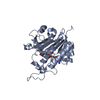
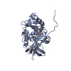
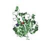
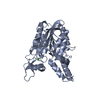
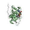
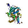
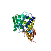
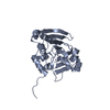
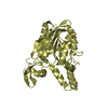
| Title | X-ray Structure of Gene Product from Arabidopsis Thaliana At5g02240 | ||||||
 Components Components | unknown protein | ||||||
 Keywords Keywords |  STRUCTURAL GENOMICS / UNKNOWN FUNCTION / PROTEIN STRUCTURE INITIATIVE / CESG / AT5G02240 / NADP / PSI / Center for Eukaryotic Structural Genomics STRUCTURAL GENOMICS / UNKNOWN FUNCTION / PROTEIN STRUCTURE INITIATIVE / CESG / AT5G02240 / NADP / PSI / Center for Eukaryotic Structural Genomics | ||||||
| Function / homology |  Function and homology information Function and homology informationresponse to abscisic acid / apoplast / plant-type vacuole / chloroplast stroma / oxidoreductase activity / plasma membrane / cytosolSimilarity search - Function | ||||||
| Biological species |  Arabidopsis thaliana (thale cress) Arabidopsis thaliana (thale cress) | ||||||
| Method | X-RAY DIFFRACTION / SYNCHROTRON / MAD / Resolution: 1.8 Å | ||||||
 Authors Authors | Wesenberg, G.E. / Smith, D.W. / Phillips Jr., G.N. / Bitto, E. / Bingman, C.A. / Allard, S.T.M. / Center for Eukaryotic Structural Genomics (CESG) | ||||||
 Citation Citation | Journal: To be published Title: X-ray Structure of Gene Product from Arabidopsis Thaliana At5g02240 Authors: Center for Eukaryotic Structural Genomics | ||||||
| History |
|
- Structure visualization
| Structure viewer | Molecule: MolmilJmol/JSmol |
|---|
- Downloads & links
-Download
| PDBx/mmCIF format | 1xq6.cif.gz | 118.7 KB | Display | PDBx/mmCIF format |
|---|---|---|---|---|
| PDB format | pdb1xq6.ent.gz | 91.3 KB | Display | PDB format |
| PDBx/mmJSON format | 1xq6.json.gz | Tree view | PDBx/mmJSON format | |
| Others |  Other downloads Other downloads |
-Validation report
| Arichive directory | https://data.pdbj.org/pub/pdb/validation_reports/xq/1xq6ftp://data.pdbj.org/pub/pdb/validation_reports/xq/1xq6 | HTTPS FTP |
|---|
-Related structure data
| Similar structure data | |
|---|---|
| Other databases |

-Links
 PDBj
PDBj- Assembly
| Deposited unit | 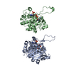
| ||||||||
|---|---|---|---|---|---|---|---|---|---|
| 1 | 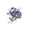
| ||||||||
| 2 | 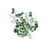
| ||||||||
| Unit cell |
|
-Components
| #1: Protein | Mass: 27092.766 Da / Num. of mol.: 2 Source method: isolated from a genetically manipulated source Source: (gene. exp.) Arabidopsis thaliana (thale cress) / Gene: At5g02240 / Plasmid: pVP-13 (pQE derivative) / Production host:  Escherichia coli (E. coli) / Strain (production host): B834(DE3) p(LacI+RARE) / References: UniProt: Q94EG6 Escherichia coli (E. coli) / Strain (production host): B834(DE3) p(LacI+RARE) / References: UniProt: Q94EG6#2: Chemical | Nicotinamide adenine dinucleotide phosphate  Mass: 743.405 Da / Num. of mol.: 2 / Source method: obtained synthetically / Formula: C21H28N7O17P3 Mass: 743.405 Da / Num. of mol.: 2 / Source method: obtained synthetically / Formula: C21H28N7O17P3#3: Water | ChemComp-HOH / | Water Mass: 18.015 Da / Num. of mol.: 400 / Source method: isolated from a natural source / Formula: H2O Mass: 18.015 Da / Num. of mol.: 400 / Source method: isolated from a natural source / Formula: H2O |
|---|
-Experimental details
-Experiment
| Experiment | Method: X-RAY DIFFRACTION / Number of used crystals: 2 |
|---|
- Sample preparation
| Crystal |
| |||||||||||||||
|---|---|---|---|---|---|---|---|---|---|---|---|---|---|---|---|---|
| Crystal grow |
|
-Data collection
| Diffraction |
| |||||||||||||||||||||||||||||||||||||||||||||||||||||||||||||||||||||||||||||
|---|---|---|---|---|---|---|---|---|---|---|---|---|---|---|---|---|---|---|---|---|---|---|---|---|---|---|---|---|---|---|---|---|---|---|---|---|---|---|---|---|---|---|---|---|---|---|---|---|---|---|---|---|---|---|---|---|---|---|---|---|---|---|---|---|---|---|---|---|---|---|---|---|---|---|---|---|---|---|
| Diffraction source |
| |||||||||||||||||||||||||||||||||||||||||||||||||||||||||||||||||||||||||||||
| Detector |
| |||||||||||||||||||||||||||||||||||||||||||||||||||||||||||||||||||||||||||||
| Radiation |
| |||||||||||||||||||||||||||||||||||||||||||||||||||||||||||||||||||||||||||||
| Radiation wavelength |
| |||||||||||||||||||||||||||||||||||||||||||||||||||||||||||||||||||||||||||||
| Reflection | Resolution: 1.8→23.565 Å / Num. obs: 52253 / % possible obs: 93.9 % / Redundancy: 5.2 % / Rmerge(I) obs: 0.043 / Χ2: 0.921 / Net I/σ(I): 19.7 | |||||||||||||||||||||||||||||||||||||||||||||||||||||||||||||||||||||||||||||
| Reflection shell |
|

-Phasing
| Phasing | Method: MAD | |||||||||||||||||||||||||||||||||||||||||||||||||
|---|---|---|---|---|---|---|---|---|---|---|---|---|---|---|---|---|---|---|---|---|---|---|---|---|---|---|---|---|---|---|---|---|---|---|---|---|---|---|---|---|---|---|---|---|---|---|---|---|---|---|
| Phasing set |
| |||||||||||||||||||||||||||||||||||||||||||||||||
| Phasing MAD | D res high: 2.05 Å / D res low: 20 Å / FOM : 0.46 / Reflection: 32618 | |||||||||||||||||||||||||||||||||||||||||||||||||
| Phasing MAD set |
| |||||||||||||||||||||||||||||||||||||||||||||||||
| Phasing MAD set site |
| |||||||||||||||||||||||||||||||||||||||||||||||||
| Phasing MAD shell |
| |||||||||||||||||||||||||||||||||||||||||||||||||
| Phasing dm | FOM : 0.61 / FOM acentric: 0.6 / FOM centric: 0.64 / Reflection: 32619 / Reflection acentric: 29470 / Reflection centric: 3149 | |||||||||||||||||||||||||||||||||||||||||||||||||
| Phasing dm shell |
|
- Processing
| Software |
| |||||||||||||||||||||||||||||||||||||||||||||||||||||||||||||||||||||||||||||||||||||||||||||||||||||||||||||||||||||||||||||||||||||||||||||||||||
|---|---|---|---|---|---|---|---|---|---|---|---|---|---|---|---|---|---|---|---|---|---|---|---|---|---|---|---|---|---|---|---|---|---|---|---|---|---|---|---|---|---|---|---|---|---|---|---|---|---|---|---|---|---|---|---|---|---|---|---|---|---|---|---|---|---|---|---|---|---|---|---|---|---|---|---|---|---|---|---|---|---|---|---|---|---|---|---|---|---|---|---|---|---|---|---|---|---|---|---|---|---|---|---|---|---|---|---|---|---|---|---|---|---|---|---|---|---|---|---|---|---|---|---|---|---|---|---|---|---|---|---|---|---|---|---|---|---|---|---|---|---|---|---|---|---|---|---|---|
| Refinement | Method to determine structure: MAD / Resolution: 1.8→23.57 Å / Cor.coef. Fo:Fc: 0.945 / Cor.coef. Fo:Fc free: 0.903 / WRfactor Rfree: 0.274 / WRfactor Rwork: 0.218 / SU B: 5.126 / SU ML: 0.154 / SU R Cruickshank DPI: 0.161 / Cross valid method: THROUGHOUT / ESU R: 0.161 / ESU R Free: 0.16 / Details: HYDROGENS HAVE BEEN ADDED IN THE RIDING POSITIONS
| |||||||||||||||||||||||||||||||||||||||||||||||||||||||||||||||||||||||||||||||||||||||||||||||||||||||||||||||||||||||||||||||||||||||||||||||||||
| Solvent computation | Ion probe radii: 0.8 Å / Shrinkage radii: 0.8 Å / VDW probe radii: 1.2 Å / Solvent model: BABINET MODEL PLUS MASK | |||||||||||||||||||||||||||||||||||||||||||||||||||||||||||||||||||||||||||||||||||||||||||||||||||||||||||||||||||||||||||||||||||||||||||||||||||
| Displacement parameters | Biso mean: 32.229 Å2
| |||||||||||||||||||||||||||||||||||||||||||||||||||||||||||||||||||||||||||||||||||||||||||||||||||||||||||||||||||||||||||||||||||||||||||||||||||
| Refinement step | Cycle: LAST / Resolution: 1.8→23.57 Å
| |||||||||||||||||||||||||||||||||||||||||||||||||||||||||||||||||||||||||||||||||||||||||||||||||||||||||||||||||||||||||||||||||||||||||||||||||||
| Refine LS restraints |
| |||||||||||||||||||||||||||||||||||||||||||||||||||||||||||||||||||||||||||||||||||||||||||||||||||||||||||||||||||||||||||||||||||||||||||||||||||
| LS refinement shell | Refine-ID: X-RAY DIFFRACTION / Total num. of bins used: 20
|
