 Movie
Movie Controller

[English] 日本語
 Yorodumi
- PDB-4mch: Crystal structure of uridine phosphorylase from vibrio fischeri e... -
+ Open data

- Basic information
| Entry | Database: PDB / ID: 4mch | ||||||
|---|---|---|---|---|---|---|---|
| Title | Crystal structure of uridine phosphorylase from vibrio fischeri es114 complexed with 6-hydroxy-1-naphthoic acid, NYSGRC Target 029520. | ||||||
 Components Components | Uridine phosphorylase | ||||||
 Keywords Keywords | TRANSFERASE / STRUCTURAL GENOMICS / PROTEIN STRUCTURE INITIATIVE / NYSGRC / PSI-Biology / New York Structural Genomics Research Consortium | ||||||
| Function / homology |  Function and homology information Function and homology informationuridine catabolic process / uridine phosphorylase / uridine phosphorylase activity / purine nucleoside catabolic process / purine-nucleoside phosphorylase activity / cytosolSimilarity search - Function | ||||||
| Biological species |  Vibrio fischeri (bacteria) Vibrio fischeri (bacteria) | ||||||
| Method | X-RAY DIFFRACTION / SYNCHROTRON / MOLECULAR REPLACEMENT / molecular replacement / Resolution: 1.73 Å | ||||||
 Authors Authors | Malashkevich, V.N. / Bhosle, R. / Toro, R. / Hillerich, B. / Gizzi, A. / Garforth, S. / Kar, A. / Chan, M.K. / Lafluer, J. / Patel, H. ...Malashkevich, V.N. / Bhosle, R. / Toro, R. / Hillerich, B. / Gizzi, A. / Garforth, S. / Kar, A. / Chan, M.K. / Lafluer, J. / Patel, H. / Matikainen, B. / Chamala, S. / Lim, S. / Celikgil, A. / Villegas, G. / Evans, B. / Love, J. / Fiser, A. / Khafizov, K. / Seidel, R. / Bonanno, J.B. / Almo, S.C. / New York Structural Genomics Research Consortium (NYSGRC) | ||||||
 Citation Citation | Journal: To be Published Title: Crystal structure of uridine phosphorylase from vibrio fischeri es114 complexed with 6-hydroxy-1-naphthoic acid, NYSGRC Target 029520. Authors: Malashkevich, V.N. / Bhosle, R. / Toro, R. / Hillerich, B. / Gizzi, A. / Garforth, S. / Kar, A. / Chan, M.K. / Lafluer, J. / Patel, H. / Matikainen, B. / Chamala, S. / Lim, S. / Celikgil, A. ...Authors: Malashkevich, V.N. / Bhosle, R. / Toro, R. / Hillerich, B. / Gizzi, A. / Garforth, S. / Kar, A. / Chan, M.K. / Lafluer, J. / Patel, H. / Matikainen, B. / Chamala, S. / Lim, S. / Celikgil, A. / Villegas, G. / Evans, B. / Love, J. / Fiser, A. / Khafizov, K. / Seidel, R. / Bonanno, J.B. / Almo, S.C. | ||||||
| History |
|
- Structure visualization
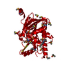
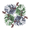
| Structure viewer | Molecule: MolmilJmol/JSmol |
|---|
- Downloads & links
-Download
| PDBx/mmCIF format | 4mch.cif.gz | 119.3 KB | Display | PDBx/mmCIF format |
|---|---|---|---|---|
| PDB format | pdb4mch.ent.gz | 91.4 KB | Display | PDB format |
| PDBx/mmJSON format | 4mch.json.gz | Tree view | PDBx/mmJSON format | |
| Others |  Other downloads Other downloads |
-Validation report
| Arichive directory | https://data.pdbj.org/pub/pdb/validation_reports/mc/4mchftp://data.pdbj.org/pub/pdb/validation_reports/mc/4mch | HTTPS FTP |
|---|
-Related structure data
| Related structure data |  4lnhS S: Starting model for refinement |
|---|---|
| Similar structure data | |
| Other databases |


-Links
 PDBj
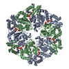
PDBj- Assembly
| Deposited unit | 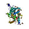
| |||||||||||||||
|---|---|---|---|---|---|---|---|---|---|---|---|---|---|---|---|---|
| 1 | x 6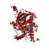
| |||||||||||||||
| Unit cell |
| |||||||||||||||
| Components on special symmetry positions |
| |||||||||||||||
| Details | hexameric |
-Components
| #1: Protein | Mass: 29829.449 Da / Num. of mol.: 1 Source method: isolated from a genetically manipulated source Source: (gene. exp.) Vibrio fischeri (bacteria) / Strain: ES114 / Gene: 3280420, VF_A0530 / Plasmid: BC-PSGX3(BC) / Production host: Escherichia coli (E. coli) / Strain (production host): BL21(DE3)CODON+RIL / References: UniProt: Q5E046, uridine phosphorylase | ||
|---|---|---|---|
| #2: Chemical | ChemComp-SO4 / Sulfate  Mass: 96.063 Da / Num. of mol.: 1 / Source method: obtained synthetically / Formula: SO4 Mass: 96.063 Da / Num. of mol.: 1 / Source method: obtained synthetically / Formula: SO4 | ||
| #3: Chemical | ChemComp-61N /   Mass: 188.179 Da / Num. of mol.: 1 / Source method: obtained synthetically / Formula: C11H8O3 Mass: 188.179 Da / Num. of mol.: 1 / Source method: obtained synthetically / Formula: C11H8O3 | ||
| #4: Chemical | ChemComp-DMS / Dimethyl sulfoxide  Mass: 78.133 Da / Num. of mol.: 14 / Source method: obtained synthetically / Formula: C2H6OS / Comment: DMSO, precipitant*YM Mass: 78.133 Da / Num. of mol.: 14 / Source method: obtained synthetically / Formula: C2H6OS / Comment: DMSO, precipitant*YM#5: Water | ChemComp-HOH / | Water Mass: 18.015 Da / Num. of mol.: 156 / Source method: isolated from a natural source / Formula: H2O Mass: 18.015 Da / Num. of mol.: 156 / Source method: isolated from a natural source / Formula: H2O |
-Experimental details
-Experiment
| Experiment | Method: X-RAY DIFFRACTION / Number of used crystals: 1 |
|---|
- Sample preparation
| Crystal | Density Matthews: 2.53 Å3/Da / Density % sol: 51.4 % |
|---|---|
| Crystal grow | Temperature: 298 K / Method: vapor diffusion, sitting drop / pH: 8.5 Details: 0.2 M AMMONIUM SULFATE, 0.1 M HEPES:NAOH, PH 8.5, 25% PEG 3350, VAPOR DIFFUSION, SITTING DROP, temperature 298K |
-Data collection
| Diffraction | Mean temperature: 100 K | |||||||||||||||||||||||||||||||||||||||||||||||||||||||||||||||||||||||||||||
|---|---|---|---|---|---|---|---|---|---|---|---|---|---|---|---|---|---|---|---|---|---|---|---|---|---|---|---|---|---|---|---|---|---|---|---|---|---|---|---|---|---|---|---|---|---|---|---|---|---|---|---|---|---|---|---|---|---|---|---|---|---|---|---|---|---|---|---|---|---|---|---|---|---|---|---|---|---|---|
| Diffraction source | Source: SYNCHROTRON / Site: APS  / Beamline: 31-ID / Wavelength: 0.9791 Å / Beamline: 31-ID / Wavelength: 0.9791 Å | |||||||||||||||||||||||||||||||||||||||||||||||||||||||||||||||||||||||||||||
| Detector | Type: RAYONIX MX225HE / Detector: CCD / Date: Aug 13, 2013 | |||||||||||||||||||||||||||||||||||||||||||||||||||||||||||||||||||||||||||||
| Radiation | Protocol: SAD / Scattering type: x-ray | |||||||||||||||||||||||||||||||||||||||||||||||||||||||||||||||||||||||||||||
| Radiation wavelength | Wavelength: 0.9791 Å / Relative weight: 1 | |||||||||||||||||||||||||||||||||||||||||||||||||||||||||||||||||||||||||||||
| Reflection | Resolution: 1.73→19.753 Å / Num. obs: 31147 / % possible obs: 99.6 % / Redundancy: 7.2 % / Rmerge(I) obs: 0.103 / Rsym value: 0.103 / Net I/σ(I): 7.9 | |||||||||||||||||||||||||||||||||||||||||||||||||||||||||||||||||||||||||||||
| Reflection shell | Rmerge(I) obs: 0.015 / Diffraction-ID: 1
|
-Phasing
| Phasing | Method: molecular replacement |
|---|
- Processing
| Software |
| ||||||||||||||||||||||||||||||||||||||||||||||||||||||||||||
|---|---|---|---|---|---|---|---|---|---|---|---|---|---|---|---|---|---|---|---|---|---|---|---|---|---|---|---|---|---|---|---|---|---|---|---|---|---|---|---|---|---|---|---|---|---|---|---|---|---|---|---|---|---|---|---|---|---|---|---|---|---|
| Refinement | Method to determine structure: MOLECULAR REPLACEMENT Starting model: PDB ENTRY 4LNH Resolution: 1.73→19.16 Å / Cor.coef. Fo:Fc: 0.967 / Cor.coef. Fo:Fc free: 0.958 / WRfactor Rfree: 0.2157 / WRfactor Rwork: 0.1871 / Occupancy max: 1 / Occupancy min: 0.5 / FOM work R set: 0.9078 / SU B: 3.152 / SU ML: 0.047 / SU R Cruickshank DPI: 0.0204 / SU Rfree: 0.0196 / Cross valid method: THROUGHOUT / σ(F): 0 / ESU R: 0.02 / ESU R Free: 0.02 / Stereochemistry target values: MAXIMUM LIKELIHOOD Details: U VALUES : WITH TLS ADDED HYDROGENS HAVE BEEN USED IF PRESENT IN THE INPUT
| ||||||||||||||||||||||||||||||||||||||||||||||||||||||||||||
| Solvent computation | Ion probe radii: 0.8 Å / Shrinkage radii: 0.8 Å / VDW probe radii: 1.2 Å / Solvent model: MASK | ||||||||||||||||||||||||||||||||||||||||||||||||||||||||||||
| Displacement parameters | Biso max: 111.25 Å2 / Biso mean: 34.5311 Å2 / Biso min: 19.47 Å2
| ||||||||||||||||||||||||||||||||||||||||||||||||||||||||||||
| Refinement step | Cycle: LAST / Resolution: 1.73→19.16 Å
| ||||||||||||||||||||||||||||||||||||||||||||||||||||||||||||
| Refine LS restraints |
| ||||||||||||||||||||||||||||||||||||||||||||||||||||||||||||
| LS refinement shell | Resolution: 1.73→1.775 Å / Total num. of bins used: 20
| ||||||||||||||||||||||||||||||||||||||||||||||||||||||||||||
| Refinement TLS params. | Method: refined / Origin x: 1.4853 Å / Origin y: 29.7345 Å / Origin z: 27.326 Å
|
