 Movie
Movie Controller

[English] 日本語
 Yorodumi
- PDB-1xdn: High resolution crystal structure of an editosome enzyme from try... -
+ Open data

- Basic information
| Entry | Database: PDB / ID: 1xdn | ||||||
|---|---|---|---|---|---|---|---|
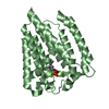
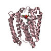
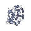
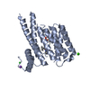
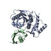
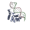
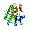
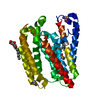
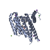
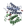
| Title | High resolution crystal structure of an editosome enzyme from trypanosoma brucei: RNA editing ligase 1 | ||||||
 Components Components | RNA editing ligase MP52 | ||||||
 Keywords Keywords |  LIGASE / RNA editing / Trypanosoma brucei LIGASE / RNA editing / Trypanosoma brucei | ||||||
| Function / homology |  Function and homology informationRNA ligase (ATP) / RNA ligase (ATP) activity / RNA modification / mitochondrial mRNA editing complex / kinetoplast / mRNA processing / mitochondrion / RNA binding / ATP binding / cytoplasm Function and homology informationRNA ligase (ATP) / RNA ligase (ATP) activity / RNA modification / mitochondrial mRNA editing complex / kinetoplast / mRNA processing / mitochondrion / RNA binding / ATP binding / cytoplasmSimilarity search - Function | ||||||
| Biological species |  Trypanosoma brucei (eukaryote) Trypanosoma brucei (eukaryote) | ||||||
| Method | X-RAY DIFFRACTION / SYNCHROTRON / MAD / Resolution: 1.2 Å | ||||||
 Authors Authors | Deng, J. / Schnaufer, A. / Salavati, R. / Stuart, K.D. / Hol, W.G. | ||||||
 Citation Citation | Journal: J.Mol.Biol. / Year: 2004 Title: High resolution crystal structure of a key editosome enzyme from Trypanosoma brucei: RNA editing ligase 1. Authors: Deng, J. / Schnaufer, A. / Salavati, R. / Stuart, K.D. / Hol, W.G. | ||||||
| History |
|
- Structure visualization
| Structure viewer | Molecule: MolmilJmol/JSmol |
|---|
- Downloads & links
-Download
| PDBx/mmCIF format | 1xdn.cif.gz | 134.9 KB | Display | PDBx/mmCIF format |
|---|---|---|---|---|
| PDB format | pdb1xdn.ent.gz | 108.8 KB | Display | PDB format |
| PDBx/mmJSON format | 1xdn.json.gz | Tree view | PDBx/mmJSON format | |
| Others |  Other downloads Other downloads |
-Validation report
| Arichive directory | https://data.pdbj.org/pub/pdb/validation_reports/xd/1xdnftp://data.pdbj.org/pub/pdb/validation_reports/xd/1xdn | HTTPS FTP |
|---|
-Related structure data
| Similar structure data |
|---|
-Links
 PDBj
PDBj- Assembly
| Deposited unit | 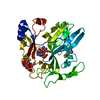
| ||||||||
|---|---|---|---|---|---|---|---|---|---|
| 1 |
| ||||||||
| Unit cell |
|
-Components
| #1: Protein | Mass: 31537.598 Da / Num. of mol.: 1 / Fragment: Adenylation domain Source method: isolated from a genetically manipulated source Source: (gene. exp.) Trypanosoma brucei (eukaryote) / Gene: MP52 / Plasmid: pskB3 / Production host:  Escherichia coli (E. coli) / Strain (production host): BL21GOLD DE3 / References: GenBank: 11067037, UniProt: P86927*PLUS Escherichia coli (E. coli) / Strain (production host): BL21GOLD DE3 / References: GenBank: 11067037, UniProt: P86927*PLUS |
|---|---|
| #2: Chemical | ChemComp-MG /   Mass: 24.305 Da / Num. of mol.: 1 / Source method: obtained synthetically / Formula: Mg Mass: 24.305 Da / Num. of mol.: 1 / Source method: obtained synthetically / Formula: Mg |
| #3: Chemical | ChemComp-ATP / Adenosine triphosphate  Mass: 507.181 Da / Num. of mol.: 1 / Source method: obtained synthetically / Formula: C10H16N5O13P3 / Comment: ATP, energy-carrying molecule*YM Mass: 507.181 Da / Num. of mol.: 1 / Source method: obtained synthetically / Formula: C10H16N5O13P3 / Comment: ATP, energy-carrying molecule*YM |
| #4: Water | ChemComp-HOH / Water Mass: 18.015 Da / Num. of mol.: 440 / Source method: isolated from a natural source / Formula: H2O Mass: 18.015 Da / Num. of mol.: 440 / Source method: isolated from a natural source / Formula: H2O |
-Experimental details
-Experiment
| Experiment | Method: X-RAY DIFFRACTION / Number of used crystals: 1 |
|---|
- Sample preparation
| Crystal | Density Matthews: 2.3 Å3/Da / Density % sol: 45.8 % |
|---|---|
| Crystal grow | Temperature: 298 K / Method: vapor diffusion, sitting drop / pH: 7.5 Details: PEG 3350, magnesium chloride, Tris, ATP, pH 7.5, VAPOR DIFFUSION, SITTING DROP, temperature 298K |
-Data collection
| Diffraction | Mean temperature: 100 K | ||||||||||||
|---|---|---|---|---|---|---|---|---|---|---|---|---|---|
| Diffraction source | Source: SYNCHROTRON / Site: APS  / Beamline: 19-ID / Wavelength: 0.97885, 0.97899, 0.96112 / Beamline: 19-ID / Wavelength: 0.97885, 0.97899, 0.96112 | ||||||||||||
| Detector | Type: ADSC QUANTUM 4 / Detector: CCD / Date: Dec 15, 2003 | ||||||||||||
| Radiation | Protocol: MAD / Monochromatic (M) / Laue (L): M / Scattering type: x-ray | ||||||||||||
| Radiation wavelength |
| ||||||||||||
| Reflection | Resolution: 1.1→50 Å / Num. obs: 95711 / % possible obs: 86.8 % / Observed criterion σ(I): 2 / Biso Wilson estimate: 7.293 Å2 / Rmerge(I) obs: 0.064 / Rsym value: 0.064 / Net I/σ(I): 33.8 | ||||||||||||
| Reflection shell | Highest resolution: 1.1 Å / Rmerge(I) obs: 0.064 / Mean I/σ(I) obs: 33.8 / Num. unique all: 95711 / Rsym value: 0.064 / % possible all: 86.8 |
- Processing
| Software |
| ||||||||||||||||||||||||||||||||||||||||||||||||||||||||||||||||||||||||||||||||||||||||||||||||||||||||||||||||||||||||||||||||||||||||||||||||||||||||||||||||
|---|---|---|---|---|---|---|---|---|---|---|---|---|---|---|---|---|---|---|---|---|---|---|---|---|---|---|---|---|---|---|---|---|---|---|---|---|---|---|---|---|---|---|---|---|---|---|---|---|---|---|---|---|---|---|---|---|---|---|---|---|---|---|---|---|---|---|---|---|---|---|---|---|---|---|---|---|---|---|---|---|---|---|---|---|---|---|---|---|---|---|---|---|---|---|---|---|---|---|---|---|---|---|---|---|---|---|---|---|---|---|---|---|---|---|---|---|---|---|---|---|---|---|---|---|---|---|---|---|---|---|---|---|---|---|---|---|---|---|---|---|---|---|---|---|---|---|---|---|---|---|---|---|---|---|---|---|---|---|---|---|---|
| Refinement | Method to determine structure: MAD / Resolution: 1.2→20 Å / Cor.coef. Fo:Fc: 0.975 / Cor.coef. Fo:Fc free: 0.972 / SU B: 0.349 / SU ML: 0.017 / Cross valid method: THROUGHOUT / ESU R: 0.034 / ESU R Free: 0.033 / Stereochemistry target values: MAXIMUM LIKELIHOOD / Details: HYDROGENS HAVE BEEN ADDED IN THE RIDING POSITIONS
| ||||||||||||||||||||||||||||||||||||||||||||||||||||||||||||||||||||||||||||||||||||||||||||||||||||||||||||||||||||||||||||||||||||||||||||||||||||||||||||||||
| Solvent computation | Ion probe radii: 0.8 Å / Shrinkage radii: 0.8 Å / VDW probe radii: 1.4 Å / Solvent model: BABINET MODEL WITH MASK | ||||||||||||||||||||||||||||||||||||||||||||||||||||||||||||||||||||||||||||||||||||||||||||||||||||||||||||||||||||||||||||||||||||||||||||||||||||||||||||||||
| Displacement parameters | Biso mean: 9.403 Å2
| ||||||||||||||||||||||||||||||||||||||||||||||||||||||||||||||||||||||||||||||||||||||||||||||||||||||||||||||||||||||||||||||||||||||||||||||||||||||||||||||||
| Refinement step | Cycle: LAST / Resolution: 1.2→20 Å
| ||||||||||||||||||||||||||||||||||||||||||||||||||||||||||||||||||||||||||||||||||||||||||||||||||||||||||||||||||||||||||||||||||||||||||||||||||||||||||||||||
| Refine LS restraints |
| ||||||||||||||||||||||||||||||||||||||||||||||||||||||||||||||||||||||||||||||||||||||||||||||||||||||||||||||||||||||||||||||||||||||||||||||||||||||||||||||||
| LS refinement shell | Resolution: 1.2→1.231 Å / Total num. of bins used: 20 /
|
