 Movie
Movie Controller

[English] 日本語
 Yorodumi
- PDB-1euv: X-RAY STRUCTURE OF THE C-TERMINAL ULP1 PROTEASE DOMAIN IN COMPLEX... -
+ Open data

- Basic information
| Entry | Database: PDB / ID: 1euv | ||||||
|---|---|---|---|---|---|---|---|
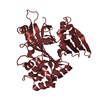
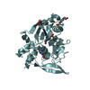
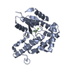
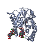
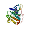
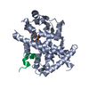
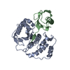
| Title | X-RAY STRUCTURE OF THE C-TERMINAL ULP1 PROTEASE DOMAIN IN COMPLEX WITH SMT3, THE YEAST ORTHOLOG OF SUMO. | ||||||
 Components Components |
| ||||||
 Keywords Keywords |  HYDROLASE / SUMO HYDROLASE / UBIQUITIN-LIKE PROTEASE 1 / SMT3 HYDROLASE DESUMOYLATING ENZYME / CYSTEINE PROTEASE / SUMO PROCESSING ENZYME / SMT3 PROCESSING ENZYME / NABH4 / THIOHEMIACETAL / COVALENT PROTEASE ADDUCT HYDROLASE / SUMO HYDROLASE / UBIQUITIN-LIKE PROTEASE 1 / SMT3 HYDROLASE DESUMOYLATING ENZYME / CYSTEINE PROTEASE / SUMO PROCESSING ENZYME / SMT3 PROCESSING ENZYME / NABH4 / THIOHEMIACETAL / COVALENT PROTEASE ADDUCT | ||||||
| Function / homology |  Function and homology informationUlp1 peptidase / SUMO is conjugated to E1 (UBA2:SAE1) / SUMOylation of nuclear envelope proteins / SUMO is transferred from E1 to E2 (UBE2I, UBC9) / SUMO is proteolytically processed / SUMOylation of transcription factors / Postmitotic nuclear pore complex (NPC) reformation / SUMOylation of transcription cofactors / deSUMOylase activity / protein desumoylation ...Ulp1 peptidase / SUMO is conjugated to E1 (UBA2:SAE1) / SUMOylation of nuclear envelope proteins / SUMO is transferred from E1 to E2 (UBE2I, UBC9) / SUMO is proteolytically processed / SUMOylation of transcription factors / Postmitotic nuclear pore complex (NPC) reformation / SUMOylation of transcription cofactors / deSUMOylase activity / protein desumoylation / SUMOylation of DNA damage response and repair proteins / SUMOylation of DNA replication proteins / septin ring / Recruitment and ATM-mediated phosphorylation of repair and signaling proteins at DNA double strand breaks / SUMOylation of SUMOylation proteins / SUMOylation of RNA binding proteins / SUMOylation of chromatin organization proteins / Major pathway of rRNA processing in the nucleolus and cytosol / ubiquitin-like protein ligase binding / protein sumoylation / cysteine-type peptidase activity / condensed nuclear chromosome / PML body / protein tag activity / G2/M transition of mitotic cell cycle / nuclear envelope / protein-containing complex binding / nucleolus / proteolysis / identical protein binding / nucleus Function and homology informationUlp1 peptidase / SUMO is conjugated to E1 (UBA2:SAE1) / SUMOylation of nuclear envelope proteins / SUMO is transferred from E1 to E2 (UBE2I, UBC9) / SUMO is proteolytically processed / SUMOylation of transcription factors / Postmitotic nuclear pore complex (NPC) reformation / SUMOylation of transcription cofactors / deSUMOylase activity / protein desumoylation ...Ulp1 peptidase / SUMO is conjugated to E1 (UBA2:SAE1) / SUMOylation of nuclear envelope proteins / SUMO is transferred from E1 to E2 (UBE2I, UBC9) / SUMO is proteolytically processed / SUMOylation of transcription factors / Postmitotic nuclear pore complex (NPC) reformation / SUMOylation of transcription cofactors / deSUMOylase activity / protein desumoylation / SUMOylation of DNA damage response and repair proteins / SUMOylation of DNA replication proteins / septin ring / Recruitment and ATM-mediated phosphorylation of repair and signaling proteins at DNA double strand breaks / SUMOylation of SUMOylation proteins / SUMOylation of RNA binding proteins / SUMOylation of chromatin organization proteins / Major pathway of rRNA processing in the nucleolus and cytosol / ubiquitin-like protein ligase binding / protein sumoylation / cysteine-type peptidase activity / condensed nuclear chromosome / PML body / protein tag activity / G2/M transition of mitotic cell cycle / nuclear envelope / protein-containing complex binding / nucleolus / proteolysis / identical protein binding / nucleusSimilarity search - Function | ||||||
| Biological species |  Saccharomyces cerevisiae (brewer's yeast) Saccharomyces cerevisiae (brewer's yeast) | ||||||
| Method | X-RAY DIFFRACTION / SYNCHROTRON / Resolution: 1.6 Å | ||||||
 Authors Authors | Mossessova, E. / Lima, C.D. | ||||||
 Citation Citation | Journal: Mol.Cell / Year: 2000 Title: Ulp1-SUMO crystal structure and genetic analysis reveal conserved interactions and a regulatory element essential for cell growth in yeast. Authors: Mossessova, E. / Lima, C.D. #1: Journal: Nature / Year: 1999Title: A New Protease Required for Cell-cycle Progression in Yeast. Authors: Li, S.J. / Hochstrasser, M. | ||||||
| History |
|
- Structure visualization
| Structure viewer | Molecule: MolmilJmol/JSmol |
|---|
- Downloads & links
-Download
| PDBx/mmCIF format | 1euv.cif.gz | 144.8 KB | Display | PDBx/mmCIF format |
|---|---|---|---|---|
| PDB format | pdb1euv.ent.gz | 117.6 KB | Display | PDB format |
| PDBx/mmJSON format | 1euv.json.gz | Tree view | PDBx/mmJSON format | |
| Others |  Other downloads Other downloads |
-Validation report
| Arichive directory | https://data.pdbj.org/pub/pdb/validation_reports/eu/1euvftp://data.pdbj.org/pub/pdb/validation_reports/eu/1euv | HTTPS FTP |
|---|
-Related structure data
| Similar structure data |
|---|


-Links
 PDBj
PDBj


- Assembly
| Deposited unit | 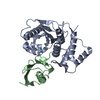
| ||||||||
|---|---|---|---|---|---|---|---|---|---|
| 1 |
| ||||||||
| Unit cell |
| ||||||||
| Details | The biological assembly is a monomer constructed from chain A. / The biological assembly is a monomer constructed from chain B. |
-Components
| #1: Protein | Ulp1 peptidase Mass: 25650.301 Da / Num. of mol.: 1 / Fragment: C-TERMINAL PROTEASE DOMAIN Source method: isolated from a genetically manipulated source Source: (gene. exp.) Saccharomyces cerevisiae (brewer's yeast)Plasmid: PET28B / Production host:  Escherichia coli (E. coli) / References: UniProt: Q02724 Escherichia coli (E. coli) / References: UniProt: Q02724 |
|---|---|
| #2: Protein | Mass: 9961.278 Da / Num. of mol.: 1 / Fragment: SMT3 RESIDUES 13-98 Source method: isolated from a genetically manipulated source Source: (gene. exp.) Saccharomyces cerevisiae (brewer's yeast)Plasmid: PET28B / Production host: Escherichia coli (E. coli) / References: UniProt: Q12306 |
| #3: Water | ChemComp-HOH / Water Mass: 18.015 Da / Num. of mol.: 432 / Source method: isolated from a natural source / Formula: H2O Mass: 18.015 Da / Num. of mol.: 432 / Source method: isolated from a natural source / Formula: H2O |
| Compound details | Covalent adduct formed between the proteolytic active site thiol and the C-terminal glycine of Smt3 ...Covalent adduct formed between the proteolytic active site thiol and the C-terminal glycine of Smt3 using the reducing agent NaBH4. |
-Experimental details
-Experiment
| Experiment | Method: X-RAY DIFFRACTION / Number of used crystals: 1 |
|---|
- Sample preparation
| Crystal | Density Matthews: 2.55 Å3/Da / Density % sol: 51.7 % | ||||||||||||||||||||
|---|---|---|---|---|---|---|---|---|---|---|---|---|---|---|---|---|---|---|---|---|---|
| Crystal grow | Temperature: 294 K / Method: vapor diffusion, hanging drop / pH: 6.5 Details: 0.1M MES pH6.5, 10% w/v polyethylene glycol 20000, 3% w/v 1,6-hexandiol, VAPOR DIFFUSION, HANGING DROP, temperature 294K | ||||||||||||||||||||
| Crystal grow | *PLUS Temperature: 221 ℃ | ||||||||||||||||||||
| Components of the solutions | *PLUS
|
-Data collection
| Diffraction | Mean temperature: 100 K |
|---|---|
| Diffraction source | Source: SYNCHROTRON / Site: NSLS  / Beamline: X4A / Wavelength: 0.9791 / Beamline: X4A / Wavelength: 0.9791 |
| Detector | Type: ADSC QUANTUM 4 / Detector: CCD / Date: Oct 21, 1999 |
| Radiation | Protocol: SINGLE WAVELENGTH / Monochromatic (M) / Laue (L): M / Scattering type: x-ray |
| Radiation wavelength | Wavelength: 0.9791 Å / Relative weight: 1 |
| Reflection | Resolution: 1.6→25 Å / Num. all: 49560 / Num. obs: 47875 / % possible obs: 96.6 % / Observed criterion σ(F): 0 / Observed criterion σ(I): 0 / Redundancy: 9.4 % / Biso Wilson estimate: 20.694 Å2 / Rmerge(I) obs: 0.043 / Net I/σ(I): 16.4 |
| Reflection shell | Resolution: 1.6→1.66 Å / Redundancy: 4.3 % / Rmerge(I) obs: 0.29 / Num. unique all: 4227 / % possible all: 86 |
| Reflection | *PLUS Num. measured all: 449291 |
| Reflection shell | *PLUS % possible obs: 86 % / Mean I/σ(I) obs: 3.4 |
- Processing
| Software |
| |||||||||||||||||||||||||
|---|---|---|---|---|---|---|---|---|---|---|---|---|---|---|---|---|---|---|---|---|---|---|---|---|---|---|
| Refinement | Resolution: 1.6→25 Å / σ(F): 1 / σ(I): 0 / Stereochemistry target values: Engh & Huber Details: Used a -loglikelihood residual derived from Rice distribution for centric and acentric cases of Fs sparse matrix procedure with anisotropic B-factor refinement.
| |||||||||||||||||||||||||
| Refinement step | Cycle: LAST / Resolution: 1.6→25 Å
| |||||||||||||||||||||||||
| Refine LS restraints |
|
