 Movie
Movie Controller

[English] 日本語
 Yorodumi
- PDB-1wtp: Hyperthermophile chromosomal protein SAC7D single mutant M29F in ... -
+ Open data

- Basic information
| Entry | Database: PDB / ID: 1wtp | ||||||
|---|---|---|---|---|---|---|---|
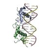
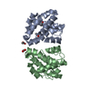
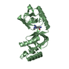
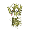
| Title | Hyperthermophile chromosomal protein SAC7D single mutant M29F in complex with DNA GCGA(UBr)CGC | ||||||
 Components Components |
| ||||||
 Keywords Keywords | DNA BINDING PROTEIN/DNA / COMPLEX CHROMATIN PROTEIN-DNA / MINOR-GROOVE DNA BINDING / ARCHEA / KINKED-DNA / INTERCALATION / Sac7d mutant / DNA BINDING PROTEIN-DNA COMPLEX | ||||||
| Function / homology |  Function and homology information Function and homology information | ||||||
| Biological species |    Sulfolobus acidocaldarius (acidophilic) Sulfolobus acidocaldarius (acidophilic) | ||||||
| Method | X-RAY DIFFRACTION / MOLECULAR REPLACEMENT / Resolution: 1.9 Å | ||||||
 Authors Authors | Chen, C.-Y. / Ko, T.-P. / Lin, T.-W. / Chou, C.-C. / Chen, C.-J. / Wang, A.H.-J. | ||||||
 Citation Citation | Journal: NUCLEIC ACIDS RES. / Year: 2005 Title: Probing the DNA kink structure induced by the hyperthermophilic chromosomal protein Sac7d Authors: Chen, C.-Y. / Ko, T.-P. / Lin, T.-W. / Chou, C.-C. / Chen, C.-J. / Wang, A.H.-J. | ||||||
| History |
|
- Structure visualization
| Structure viewer | Molecule: MolmilJmol/JSmol |
|---|
- Downloads & links
-Download
| PDBx/mmCIF format | 1wtp.cif.gz | 61.2 KB | Display | PDBx/mmCIF format |
|---|---|---|---|---|
| PDB format | pdb1wtp.ent.gz | 41.9 KB | Display | PDB format |
| PDBx/mmJSON format | 1wtp.json.gz | Tree view | PDBx/mmJSON format | |
| Others |  Other downloads Other downloads |
-Validation report
| Arichive directory | https://data.pdbj.org/pub/pdb/validation_reports/wt/1wtpftp://data.pdbj.org/pub/pdb/validation_reports/wt/1wtp | HTTPS FTP |
|---|
-Related structure data
| Related structure data | 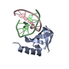 1wtoC  1wtqC  1wtrC 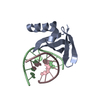 1wtvC 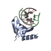 1wtwC  1wtxC 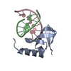 1xyiC  1azpS S: Starting model for refinement C: citing same article ( |
|---|---|
| Similar structure data |


-Links
 PDBj
PDBj- Assembly
| Deposited unit | 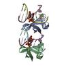
| ||||||||
|---|---|---|---|---|---|---|---|---|---|
| 1 |
| ||||||||
| Unit cell |
|
-Components
| #1: DNA chain | Mass: 2492.475 Da / Num. of mol.: 4 / Source method: obtained synthetically #2: Protein | Mass: 7642.892 Da / Num. of mol.: 2 / Mutation: M29F Source method: isolated from a genetically manipulated source Source: (gene. exp.) Sulfolobus acidocaldarius (acidophilic)Plasmid: pET3B / Production host:  Escherichia coli (E. coli) / Strain (production host): BL21(DE3)pLysS / References: UniProt: P13123 Escherichia coli (E. coli) / Strain (production host): BL21(DE3)pLysS / References: UniProt: P13123#3: Water | ChemComp-HOH / | Water Mass: 18.015 Da / Num. of mol.: 156 / Source method: isolated from a natural source / Formula: H2O Mass: 18.015 Da / Num. of mol.: 156 / Source method: isolated from a natural source / Formula: H2O |
|---|
-Experimental details
-Experiment
| Experiment | Method: X-RAY DIFFRACTION / Number of used crystals: 1 |
|---|
- Sample preparation
| Crystal | Density Matthews: 1.89 Å3/Da / Density % sol: 35 % | ||||||||||||||||||||
|---|---|---|---|---|---|---|---|---|---|---|---|---|---|---|---|---|---|---|---|---|---|
| Crystal grow | Temperature: 298 K / Method: vapor diffusion, sitting drop / pH: 7 Details: PEG 400, Tris buffer, pH 7.0, VAPOR DIFFUSION, SITTING DROP, temperature 298.0K | ||||||||||||||||||||
| Components of the solutions |
|
-Data collection
| Diffraction | Mean temperature: 150 K |
|---|---|
| Diffraction source | Source: ROTATING ANODE / Type: RIGAKU MICROMAX-002 / Wavelength: 1.5418 Å |
| Detector | Type: RIGAKU RAXIS IV++ / Detector: IMAGE PLATE / Date: Sep 16, 2002 |
| Radiation | Monochromator: GRAPHITE / Protocol: SINGLE WAVELENGTH / Monochromatic (M) / Laue (L): M / Scattering type: x-ray |
| Radiation wavelength | Wavelength: 1.5418 Å / Relative weight: 1 |
| Reflection | Resolution: 1.9→40 Å / Num. all: 15060 / Num. obs: 14849 / % possible obs: 98.6 % / Observed criterion σ(F): 2 / Observed criterion σ(I): 1 / Redundancy: 3.7 % / Rmerge(I) obs: 0.049 / Net I/σ(I): 26.4 |
| Reflection shell | Resolution: 1.9→1.97 Å / Redundancy: 2.7 % / Rmerge(I) obs: 0.269 / Mean I/σ(I) obs: 4 / Num. unique all: 1494 / % possible all: 93.4 |
- Processing
| Software |
| |||||||||||||||||||||||||
|---|---|---|---|---|---|---|---|---|---|---|---|---|---|---|---|---|---|---|---|---|---|---|---|---|---|---|
| Refinement | Method to determine structure: MOLECULAR REPLACEMENT Starting model: 1AZP Resolution: 1.9→40 Å / Cross valid method: THROUGHOUT / σ(F): 0 / Stereochemistry target values: Engh & Huber
| |||||||||||||||||||||||||
| Refine analyze |
| |||||||||||||||||||||||||
| Refinement step | Cycle: LAST / Resolution: 1.9→40 Å
| |||||||||||||||||||||||||
| Refine LS restraints |
| |||||||||||||||||||||||||
| LS refinement shell | Resolution: 1.9→1.97 Å / Rfactor Rfree error: 0.044
|
