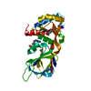
Rb-E2F complex / lens fiber cell apoptotic process / negative regulation of fat cell proliferation / regulation of cell cycle process / miRNA catabolic process / RISC complex binding / Inhibition of replication initiation of damaged DNA by RB1/E2F1 / nuclease activity / dense body / Transcription of E2F targets under negative control by p107 (RBL1) and p130 (RBL2) in complex with HDAC1 ...Rb-E2F complex / lens fiber cell apoptotic process / negative regulation of fat cell proliferation / regulation of cell cycle process / miRNA catabolic process / RISC complex binding / Inhibition of replication initiation of damaged DNA by RB1/E2F1 / nuclease activity / dense body / Transcription of E2F targets under negative control by p107 (RBL1) and p130 (RBL2) in complex with HDAC1 / mRNA stabilization / Transcription of E2F targets under negative control by DREAM complex / regulatory ncRNA-mediated gene silencing / Activation of NOXA and translocation to mitochondria / anoikis / RISC complex / endonuclease activity, active with either ribo- or deoxyribonucleic acids and producing 3'-phosphomonoesters / micrococcal nuclease / Activation of PUMA and translocation to mitochondria / DNA-binding transcription activator activity / negative regulation of fat cell differentiation / G1/S-Specific Transcription / G2 Phase / Transcriptional Regulation by E2F6 / negative regulation of DNA binding / regulation of G1/S transition of mitotic cell cycle / mRNA catabolic process / Defective binding of RB1 mutants to E2F1,(E2F2, E2F3) / intrinsic apoptotic signaling pathway by p53 class mediator / cis-regulatory region sequence-specific DNA binding / TP53 Regulates Transcription of Genes Involved in G1 Cell Cycle Arrest / Cyclin E associated events during G1/S transition / Cyclin A:Cdk2-associated events at S phase entry / forebrain development / RNA endonuclease activity / DNA damage checkpoint signaling / transcription coregulator activity / Oncogene Induced Senescence / G1/S transition of mitotic cell cycle / Pre-NOTCH Transcription and Translation / osteoblast differentiation / Transcriptional regulation of granulopoiesis / RNA polymerase II transcription regulator complex / Cyclin D associated events in G1 / intrinsic apoptotic signaling pathway in response to DNA damage / positive regulation of fibroblast proliferation / Signaling by BRAF and RAF1 fusions / sequence-specific double-stranded DNA binding / melanosome / cellular response to xenobiotic stimulus / endonuclease activity / Oxidative Stress Induced Senescence / DNA-binding transcription factor binding / sequence-specific DNA binding / molecular adaptor activity / protein dimerization activity / DNA-binding transcription factor activity, RNA polymerase II-specific / cadherin binding / positive regulation of apoptotic process / RNA polymerase II cis-regulatory region sequence-specific DNA binding / DNA-binding transcription factor activity / negative regulation of DNA-templated transcription / centrosome / DNA-templated transcription / chromatin / regulation of DNA-templated transcription / positive regulation of gene expression / regulation of transcription by RNA polymerase II / positive regulation of DNA-templated transcription / negative regulation of transcription by RNA polymerase II / positive regulation of transcription by RNA polymerase II / protein-containing complex / DNA binding / RNA binding / extracellular exosome / nucleoplasm / membrane / nucleus / cytosol / cytoplasm Similarity search - Function
Resolution: 1.45→28.14 Å / Cor.coef. Fo:Fc: 0.95 / Cor.coef. Fo:Fc free: 0.935 / SU B: 1.615 / SU ML: 0.063 / Cross valid method: THROUGHOUT / ESU R: 0.089 / ESU R Free: 0.09 / Details: HYDROGENS HAVE BEEN ADDED IN THE RIDING POSITIONS
Rfactor
Num. reflection
% reflection
Selection details
Rfree
0.2612
1920
5 %
RANDOM
Rwork
0.22892
-
-
-
obs
0.23039
36421
95.83 %
-
Solvent computation
Ion probe radii: 0.8 Å / Shrinkage radii: 0.8 Å / VDW probe radii: 1.2 Å
 Movie
Movie Controller

 Yorodumi
 Open data
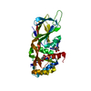
 Basic information
 Components
 Keywords
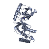
 TRANSCRIPTION / Transcriptional coactivator /
  Function and homology information
 Authors
 Citation
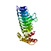
 Structure visualization
 Downloads & links
 Other downloads


 PDBj
PDBj


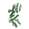
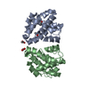
 Assembly
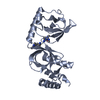

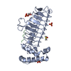
 Mass: 18.015 Da / Num. of mol.: 166 / Source method: isolated from a natural source / Formula: H2O
 Sample preparation
 / Beamline: I04 / Wavelength: 0.9795 Å
 Processing