 Movie
Movie Controller


+ Open data

- Basic information
| Entry | Database: PDB / ID: 1waa | ||||||
|---|---|---|---|---|---|---|---|
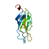
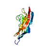
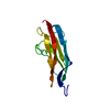
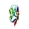
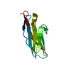
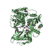
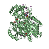
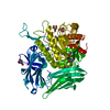
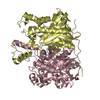
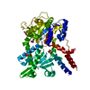
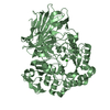
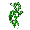
| Title | IG27 protein domain | ||||||
 Components Components | (TITIN ) x 3 ) x 3 | ||||||
 Keywords Keywords | METAL BINDING PROTEIN / CALMODULIN-BINDING / CYTOSKELETON / IMMUNOGLOBULIN DOMAIN / MUSCLE PROTEIN / PHOSPHORYLATION / SERINE/THREONINE- PROTEIN KINASE / STRUCTURAL PROTEIN. | ||||||
| Function / homology |  Function and homology information Function and homology informationsarcomerogenesis / structural molecule activity conferring elasticity / telethonin binding / skeletal muscle myosin thick filament assembly / cardiac myofibril assembly / muscle alpha-actinin binding / detection of muscle stretch / cardiac muscle tissue morphogenesis / regulation of catalytic activity / cardiac muscle hypertrophy ...sarcomerogenesis / structural molecule activity conferring elasticity / telethonin binding / skeletal muscle myosin thick filament assembly / cardiac myofibril assembly / muscle alpha-actinin binding / detection of muscle stretch / cardiac muscle tissue morphogenesis / regulation of catalytic activity / cardiac muscle hypertrophy / mitotic chromosome condensation / Striated Muscle Contraction / M band / actinin binding / I band / cardiac muscle cell development / regulation of protein kinase activity / sarcomere organization / structural constituent of muscle / skeletal muscle thin filament assembly / striated muscle thin filament / striated muscle contraction / protein kinase A signaling / cardiac muscle contraction / muscle contraction / condensed nuclear chromosome / positive regulation of protein secretion / Z disc / response to calcium ion / : / actin filament binding / Platelet degranulation / protein tyrosine kinase activity / protease binding / calmodulin binding / non-specific serine/threonine protein kinase / phosphorylation / protein serine kinase activity / protein serine/threonine kinase activity / calcium ion binding / positive regulation of gene expression / protein kinase binding / enzyme binding / extracellular exosome / extracellular region / ATP binding / identical protein binding / cytosolSimilarity search - Function | ||||||
| Biological species |  HOMO SAPIENS (human) HOMO SAPIENS (human) | ||||||
| Method | X-RAY DIFFRACTION / SYNCHROTRON / MOLECULAR REPLACEMENT / Resolution: 1.8 Å | ||||||
 Authors Authors | Vega, M.C. / Valencia, L. / Zou, P. / Wilmanns, M. | ||||||
 Citation Citation | Journal: Plos Comput.Biol. / Year: 2009 Title: Mechanical Network in Titin Immunoglobulin from Force Distribution Analysis. Authors: Stacklies, W. / Vega, M.C. / Wilmanns, M. / Grater, F. | ||||||
| History |
|
- Structure visualization
| Structure viewer | Molecule: MolmilJmol/JSmol |
|---|
- Downloads & links
-Download
| PDBx/mmCIF format | 1waa.cif.gz | 134.9 KB | Display | PDBx/mmCIF format |
|---|---|---|---|---|
| PDB format | pdb1waa.ent.gz | 105.2 KB | Display | PDB format |
| PDBx/mmJSON format | 1waa.json.gz | Tree view | PDBx/mmJSON format | |
| Others |  Other downloads Other downloads |
-Validation report
| Arichive directory | https://data.pdbj.org/pub/pdb/validation_reports/wa/1waaftp://data.pdbj.org/pub/pdb/validation_reports/wa/1waa | HTTPS FTP |
|---|
-Related structure data
| Related structure data | 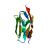 1titS S: Starting model for refinement |
|---|---|
| Similar structure data |

-Links
 PDBj
PDBj


- Assembly
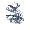
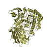
| Deposited unit | 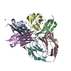
| ||||||||
|---|---|---|---|---|---|---|---|---|---|
| 1 |
| ||||||||
| Unit cell |
|
-Components
| #1: Protein | / I27 DOMAIN FROM TITIN / HEART ISOFORM N2-B Mass: 10124.597 Da / Num. of mol.: 4 / Fragment: IG DOMAIN, RESIDUES 12801-12889 Source method: isolated from a genetically manipulated source Source: (gene. exp.) HOMO SAPIENS (human) / Tissue: MUSCLESkeletal muscle / Organ: HEART / Production host:  ESCHERICHIA COLI (E. coli) ESCHERICHIA COLI (E. coli)References: UniProt: Q8WZ42, Transferases; Transferring phosphorus-containing groups; Phosphotransferases with an alcohol group as acceptor#2: Protein | | / I27 DOMAIN FROM TITIN / HEART ISOFORM N2-BMass: 10138.623 Da / Num. of mol.: 1 / Fragment: IG DOMAIN, RESIDUES 12801-12889 Source method: isolated from a genetically manipulated source Source: (gene. exp.) HOMO SAPIENS (human) / Tissue: MUSCLESkeletal muscle / Organ: HEART / Production host: ESCHERICHIA COLI (E. coli)References: UniProt: Q8WZ42, Transferases; Transferring phosphorus-containing groups; Phosphotransferases with an alcohol group as acceptor#3: Protein | | / I27 DOMAIN FROM TITIN / HEART ISOFORM N2-BMass: 10138.622 Da / Num. of mol.: 1 / Fragment: IG DOMAIN, RESIDUES 12801-12889 Source method: isolated from a genetically manipulated source Source: (gene. exp.) HOMO SAPIENS (human) / Tissue: MUSCLESkeletal muscle / Organ: HEART / Production host: ESCHERICHIA COLI (E. coli)References: UniProt: Q8WZ42, Transferases; Transferring phosphorus-containing groups; Phosphotransferases with an alcohol group as acceptor#4: Chemical | ChemComp-ZN /   Mass: 65.409 Da / Num. of mol.: 24 / Source method: obtained synthetically / Formula: Zn Mass: 65.409 Da / Num. of mol.: 24 / Source method: obtained synthetically / Formula: Zn#5: Water | ChemComp-HOH / | Water Mass: 18.015 Da / Num. of mol.: 480 / Source method: isolated from a natural source / Formula: H2O Mass: 18.015 Da / Num. of mol.: 480 / Source method: isolated from a natural source / Formula: H2OCompound details | FUNCTION: THIS MUSCLE PROTEIN MAY BE INVOLVED IN MUSCLE ASSEMBLY AND MAINTAINING THE STRUCTURAL ...FUNCTION: THIS MUSCLE PROTEIN MAY BE INVOLVED IN MUSCLE ASSEMBLY AND MAINTAININ | |
|---|
-Experimental details
-Experiment
| Experiment | Method: X-RAY DIFFRACTION |
|---|
- Sample preparation
| Crystal | Density Matthews: 2.61 Å3/Da / Density % sol: 52.9 % |
|---|
-Data collection
| Diffraction | Mean temperature: 100 K |
|---|---|
| Diffraction source | Source: SYNCHROTRON / Site: EMBL/DESY, HAMBURG  / Beamline: BW7B / Wavelength: 0.84 / Beamline: BW7B / Wavelength: 0.84 |
| Detector | Type: MARRESEARCH / Detector: CCD |
| Radiation | Protocol: SINGLE WAVELENGTH / Monochromatic (M) / Laue (L): M / Scattering type: x-ray |
| Radiation wavelength | Wavelength: 0.84 Å / Relative weight: 1 |
| Reflection | Resolution: 1.8→20 Å / Num. obs: 53298 / % possible obs: 89.2 % / Observed criterion σ(I): 0 / Redundancy: 5.5 % / Rmerge(I) obs: 0.08 / Net I/σ(I): 11.3 |
| Reflection shell | Resolution: 1.8→2 Å / Redundancy: 5.6 % / Rmerge(I) obs: 0.18 / Mean I/σ(I) obs: 5.4 / % possible all: 92.3 |
- Processing
| Software |
| ||||||||||||||||||||||||||||||||||||||||||||||||||||||||||||||||||||||||||||||||||||||||||||||||||||||||||||||||||||||||||||||||||||||||||||||||||||||||||||||||||||||||||||||||||||||
|---|---|---|---|---|---|---|---|---|---|---|---|---|---|---|---|---|---|---|---|---|---|---|---|---|---|---|---|---|---|---|---|---|---|---|---|---|---|---|---|---|---|---|---|---|---|---|---|---|---|---|---|---|---|---|---|---|---|---|---|---|---|---|---|---|---|---|---|---|---|---|---|---|---|---|---|---|---|---|---|---|---|---|---|---|---|---|---|---|---|---|---|---|---|---|---|---|---|---|---|---|---|---|---|---|---|---|---|---|---|---|---|---|---|---|---|---|---|---|---|---|---|---|---|---|---|---|---|---|---|---|---|---|---|---|---|---|---|---|---|---|---|---|---|---|---|---|---|---|---|---|---|---|---|---|---|---|---|---|---|---|---|---|---|---|---|---|---|---|---|---|---|---|---|---|---|---|---|---|---|---|---|---|---|
| Refinement | Method to determine structure: MOLECULAR REPLACEMENT Starting model: PDB ENTRY 1TIT Resolution: 1.8→15 Å / Cor.coef. Fo:Fc: 0.951 / Cor.coef. Fo:Fc free: 0.904 / SU B: 13.244 / SU ML: 0.16 / Cross valid method: THROUGHOUT / ESU R: 0.159 / ESU R Free: 0.162 / Stereochemistry target values: MAXIMUM LIKELIHOOD / Details: HYDROGENS HAVE BEEN ADDED IN THE RIDING POSITIONS.
| ||||||||||||||||||||||||||||||||||||||||||||||||||||||||||||||||||||||||||||||||||||||||||||||||||||||||||||||||||||||||||||||||||||||||||||||||||||||||||||||||||||||||||||||||||||||
| Solvent computation | Ion probe radii: 0.8 Å / Shrinkage radii: 0.8 Å / VDW probe radii: 1.2 Å / Solvent model: BABINET MODEL WITH MASK | ||||||||||||||||||||||||||||||||||||||||||||||||||||||||||||||||||||||||||||||||||||||||||||||||||||||||||||||||||||||||||||||||||||||||||||||||||||||||||||||||||||||||||||||||||||||
| Displacement parameters | Biso mean: 29.07 Å2
| ||||||||||||||||||||||||||||||||||||||||||||||||||||||||||||||||||||||||||||||||||||||||||||||||||||||||||||||||||||||||||||||||||||||||||||||||||||||||||||||||||||||||||||||||||||||
| Refinement step | Cycle: LAST / Resolution: 1.8→15 Å
| ||||||||||||||||||||||||||||||||||||||||||||||||||||||||||||||||||||||||||||||||||||||||||||||||||||||||||||||||||||||||||||||||||||||||||||||||||||||||||||||||||||||||||||||||||||||
| Refine LS restraints |
|