 Movie
Movie Controller

[English] 日本語
 Yorodumi
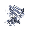
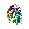
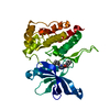
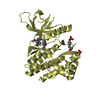
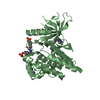
- PDB-1tki: AUTOINHIBITED SERINE KINASE DOMAIN OF THE GIANT MUSCLE PROTEIN TITIN -
+ Open data

- Basic information
| Entry | Database: PDB / ID: 1tki | ||||||
|---|---|---|---|---|---|---|---|
| Title | AUTOINHIBITED SERINE KINASE DOMAIN OF THE GIANT MUSCLE PROTEIN TITIN | ||||||
 Components Components | TITIN | ||||||
 Keywords Keywords | SERINE KINASE / TITIN / MUSCLE / AUTOINHIBITION | ||||||
| Function / homology |  Function and homology information Function and homology informationsarcomerogenesis / structural molecule activity conferring elasticity / telethonin binding / skeletal muscle myosin thick filament assembly / cardiac myofibril assembly / muscle alpha-actinin binding / detection of muscle stretch / cardiac muscle tissue morphogenesis / regulation of catalytic activity / cardiac muscle hypertrophy ...sarcomerogenesis / structural molecule activity conferring elasticity / telethonin binding / skeletal muscle myosin thick filament assembly / cardiac myofibril assembly / muscle alpha-actinin binding / detection of muscle stretch / cardiac muscle tissue morphogenesis / regulation of catalytic activity / cardiac muscle hypertrophy / mitotic chromosome condensation / Striated Muscle Contraction / actinin binding / M band / I band / cardiac muscle cell development / regulation of protein kinase activity / structural constituent of muscle / sarcomere organization / skeletal muscle thin filament assembly / striated muscle thin filament / striated muscle contraction / cardiac muscle contraction / protein kinase A signaling / condensed nuclear chromosome / muscle contraction / positive regulation of protein secretion / Z disc / response to calcium ion / : / actin filament binding / Platelet degranulation / protein tyrosine kinase activity / protease binding / calmodulin binding / non-specific serine/threonine protein kinase / phosphorylation / protein serine kinase activity / protein serine/threonine kinase activity / calcium ion binding / positive regulation of gene expression / protein kinase binding / enzyme binding / extracellular exosome / extracellular region / ATP binding / identical protein binding / cytosolSimilarity search - Function | ||||||
| Biological species |  Homo sapiens (human) Homo sapiens (human) | ||||||
| Method | X-RAY DIFFRACTION / SYNCHROTRON / molecular replacement, SINGLE CRYSTAL AVERAGING / Resolution: 2 Å | ||||||
 Authors Authors | Mayans, M.O. / Gautel, M. / Wilmanns, M. | ||||||
 Citation Citation | Journal: Nature / Year: 1998 Title: Structural basis for activation of the titin kinase domain during myofibrillogenesis. Authors: Mayans, O. / van der Ven, P.F. / Wilm, M. / Mues, A. / Young, P. / Furst, D.O. / Wilmanns, M. / Gautel, M. #1: Journal: To be PublishedTitle: X-Ray Analysis of Protein Crystals with Thin Plate Morphology Authors: Mayans, O. / Wilmanns, M. | ||||||
| History |
|
- Structure visualization
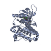
| Structure viewer | Molecule: MolmilJmol/JSmol |
|---|
- Downloads & links
-Download
| PDBx/mmCIF format | 1tki.cif.gz | 166.8 KB | Display | PDBx/mmCIF format |
|---|---|---|---|---|
| PDB format | pdb1tki.ent.gz | 118.4 KB | Display | PDB format |
| PDBx/mmJSON format | 1tki.json.gz | Tree view | PDBx/mmJSON format | |
| Others |  Other downloads Other downloads |
-Validation report
| Arichive directory | https://data.pdbj.org/pub/pdb/validation_reports/tk/1tkiftp://data.pdbj.org/pub/pdb/validation_reports/tk/1tki | HTTPS FTP |
|---|
-Related structure data
| Related structure data |  1kobS S: Starting model for refinement |
|---|---|
| Similar structure data |


-Links
 PDBj
PDBj


- Assembly
| Deposited unit | 
| ||||||||
|---|---|---|---|---|---|---|---|---|---|
| 1 | 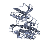
| ||||||||
| 2 | 
| ||||||||
| Unit cell |
| ||||||||
| Noncrystallographic symmetry (NCS) | NCS oper: (Code: given Matrix: (-0.999999, 0.00086, -0.001243), Vector : |
-Components
| #1: Protein | Mass: 37168.898 Da / Num. of mol.: 2 / Fragment: KINASE Source method: isolated from a genetically manipulated source Source: (gene. exp.) Homo sapiens (human) / Cell line: SF9 / Organ: HEART / Cell line (production host): SF9 / Production host:   Spodoptera frugiperda (fall armyworm) / References: UniProt: Q10466, UniProt: Q8WZ42*PLUS Spodoptera frugiperda (fall armyworm) / References: UniProt: Q10466, UniProt: Q8WZ42*PLUS#2: Water | ChemComp-HOH / | Water Mass: 18.015 Da / Num. of mol.: 514 / Source method: isolated from a natural source / Formula: H2O Mass: 18.015 Da / Num. of mol.: 514 / Source method: isolated from a natural source / Formula: H2O |
|---|
-Experimental details
-Experiment
| Experiment | Method: X-RAY DIFFRACTION / Number of used crystals: 1 |
|---|
- Sample preparation
| Crystal | Density Matthews: 2.63 Å3/Da / Density % sol: 47 % | |||||||||||||||||||||||||
|---|---|---|---|---|---|---|---|---|---|---|---|---|---|---|---|---|---|---|---|---|---|---|---|---|---|---|
| Crystal grow | *PLUS pH: 4.9 / Method: vapor diffusion | |||||||||||||||||||||||||
| Components of the solutions | *PLUS
|
-Data collection
| Diffraction | Mean temperature: 100 K |
|---|---|
| Diffraction source | Source: SYNCHROTRON / Site: EMBL/DESY, HAMBURG  / Beamline: BW7B / Wavelength: 0.84 / Beamline: BW7B / Wavelength: 0.84 |
| Detector | Type: MARRESEARCH / Detector: IMAGE PLATE / Date: Dec 1, 1997 / Details: PREMIRROR/BENT MIRROR |
| Radiation | Monochromator: GE(111) / Monochromatic (M) / Laue (L): M / Scattering type: x-ray |
| Radiation wavelength | Wavelength: 0.84 Å / Relative weight: 1 |
| Reflection | Resolution: 2→40 Å / Num. obs: 53151 / % possible obs: 96 % / Redundancy: 3.7 % / Biso Wilson estimate: 19.5 Å2 / Rsym value: 0.077 |
| Reflection shell | Resolution: 2→2.39 Å / Redundancy: 2.6 % / Rsym value: 0.239 / % possible all: 94.45 |
| Reflection | *PLUS Rmerge(I) obs: 0.077 |
| Reflection shell | *PLUS % possible obs: 94.5 % / Rmerge(I) obs: 0.239 |
- Processing
| Software |
| ||||||||||||||||||||||||||||||||||||||||||||||||||||||||||||||||||||||||||||||||
|---|---|---|---|---|---|---|---|---|---|---|---|---|---|---|---|---|---|---|---|---|---|---|---|---|---|---|---|---|---|---|---|---|---|---|---|---|---|---|---|---|---|---|---|---|---|---|---|---|---|---|---|---|---|---|---|---|---|---|---|---|---|---|---|---|---|---|---|---|---|---|---|---|---|---|---|---|---|---|---|---|---|
| Refinement | Method to determine structure: molecular replacement, SINGLE CRYSTAL AVERAGING Starting model: TRIMMED VERSION OF THE CATALYTIC DOMAIN OF TWITCHIN KINASE (1KOB) Resolution: 2→40 Å / Data cutoff high absF: 100000000000 / Data cutoff low absF: 0 / Isotropic thermal model: RESTRAINED / Cross valid method: THROUGHOUT / σ(F): 0 / Details: BULK SOLVENT CORRECTION APPLIED
| ||||||||||||||||||||||||||||||||||||||||||||||||||||||||||||||||||||||||||||||||
| Displacement parameters | Biso mean: 22.09 Å2
| ||||||||||||||||||||||||||||||||||||||||||||||||||||||||||||||||||||||||||||||||
| Refinement step | Cycle: LAST / Resolution: 2→40 Å
| ||||||||||||||||||||||||||||||||||||||||||||||||||||||||||||||||||||||||||||||||
| Refine LS restraints |
| ||||||||||||||||||||||||||||||||||||||||||||||||||||||||||||||||||||||||||||||||
| Refine LS restraints NCS | NCS model details: RESTRAINED / Weight Biso : 1 / Weight position: 300 | ||||||||||||||||||||||||||||||||||||||||||||||||||||||||||||||||||||||||||||||||
| LS refinement shell | Resolution: 2→2.28 Å
| ||||||||||||||||||||||||||||||||||||||||||||||||||||||||||||||||||||||||||||||||
| Xplor file |
| ||||||||||||||||||||||||||||||||||||||||||||||||||||||||||||||||||||||||||||||||
| Software | *PLUS Name: X-PLOR / Version: 3.1 / Classification: refinement | ||||||||||||||||||||||||||||||||||||||||||||||||||||||||||||||||||||||||||||||||
| Refinement | *PLUS | ||||||||||||||||||||||||||||||||||||||||||||||||||||||||||||||||||||||||||||||||
| Solvent computation | *PLUS | ||||||||||||||||||||||||||||||||||||||||||||||||||||||||||||||||||||||||||||||||
| Displacement parameters | *PLUS | ||||||||||||||||||||||||||||||||||||||||||||||||||||||||||||||||||||||||||||||||
| Refine LS restraints | *PLUS
| ||||||||||||||||||||||||||||||||||||||||||||||||||||||||||||||||||||||||||||||||
| LS refinement shell | *PLUS Rfactor obs: 0.224 |
