 Movie
Movie Controller

[English] 日本語
 Yorodumi
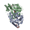
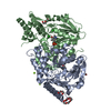
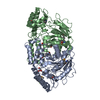
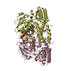
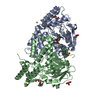
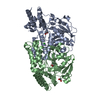
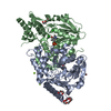
- PDB-1w3u: Crystal structure of phosphoserine aminotransferase from Bacillus... -
+ Open data

- Basic information
| Entry | Database: PDB / ID: 1w3u | ||||||
|---|---|---|---|---|---|---|---|
| Title | Crystal structure of phosphoserine aminotransferase from Bacillus circulans var. alkalophilus | ||||||
 Components Components | PHOSPHOSERINE AMINOTRANSFERASE Phosphoserine transaminase Phosphoserine transaminase | ||||||
 Keywords Keywords | TRANSFERASE / PHOSPHOSERINE AMINOTRANSFERASE / PYRIDOXAL-5'-PHOSPHATE TRANSFERASE / PYRIDINE SERINE BIOSYNTHESIS | ||||||
| Function / homology |  Function and homology informationphosphoserine transaminase / O-phospho-L-serine:2-oxoglutarate aminotransferase activity / L-serine biosynthetic process / pyridoxal phosphate binding / cytoplasm Function and homology informationphosphoserine transaminase / O-phospho-L-serine:2-oxoglutarate aminotransferase activity / L-serine biosynthetic process / pyridoxal phosphate binding / cytoplasmSimilarity search - Function | ||||||
| Biological species |  BACILLUS CIRCULANS (bacteria) BACILLUS CIRCULANS (bacteria) | ||||||
| Method | X-RAY DIFFRACTION / SYNCHROTRON / MOLECULAR REPLACEMENT / Resolution: 1.5 Å | ||||||
 Authors Authors | Kapetaniou, E.G. / Dubnovitsky, A.P. / Papageorgiou, A.C. | ||||||
 Citation Citation | Journal: Protein Sci. / Year: 2005 Title: Enzyme Adaptation to Alkaline Ph: Atomic Resolution (1.08 A) Structure of Phosphoserine Aminotransferase from Bacillus Alcalophilus Authors: Dubnovitsky, A.P. / Kapetaniou, E.G. / Papageorgiou, A.C. | ||||||
| History |
|
- Structure visualization
| Structure viewer | Molecule: MolmilJmol/JSmol |
|---|
- Downloads & links
-Download
| PDBx/mmCIF format | 1w3u.cif.gz | 125.5 KB | Display | PDBx/mmCIF format |
|---|---|---|---|---|
| PDB format | pdb1w3u.ent.gz | 95.8 KB | Display | PDB format |
| PDBx/mmJSON format | 1w3u.json.gz | Tree view | PDBx/mmJSON format | |
| Others |  Other downloads Other downloads |
-Validation report
| Arichive directory | https://data.pdbj.org/pub/pdb/validation_reports/w3/1w3uftp://data.pdbj.org/pub/pdb/validation_reports/w3/1w3u | HTTPS FTP |
|---|
-Related structure data
| Related structure data |  1w23C  1bt4S S: Starting model for refinement C: citing same article ( |
|---|---|
| Similar structure data |


-Links
 PDBj
PDBj- Assembly
| Deposited unit | 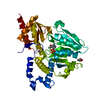
| ||||||||
|---|---|---|---|---|---|---|---|---|---|
| 1 | 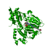
| ||||||||
| Unit cell |
|
-Components
| #1: Protein | Phosphoserine transaminase / PHOSPHOHYDROXYTHREONINE AMINOTRANSFERASE / PSAT Mass: 39966.090 Da / Num. of mol.: 1 / Mutation: YES Source method: isolated from a genetically manipulated source Details: SCHIFF BASE LINK BETWEEN A 197 AND A 363 / Source: (gene. exp.) BACILLUS CIRCULANS (bacteria) / Variant: ALKALOPHILUS / Production host: ESCHERICHIA COLI (E. coli) / Strain (production host): XL1-BLUE / References: UniProt: Q59196, phosphoserine transaminase |
|---|---|
| #2: Chemical | ChemComp-PLP / Pyridoxal phosphate  Mass: 247.142 Da / Num. of mol.: 1 / Source method: obtained synthetically / Formula: C8H10NO6P Mass: 247.142 Da / Num. of mol.: 1 / Source method: obtained synthetically / Formula: C8H10NO6P |
| #3: Chemical | ChemComp-GOL / Glycerol  Mass: 92.094 Da / Num. of mol.: 1 / Source method: obtained synthetically / Formula: C3H8O3 Mass: 92.094 Da / Num. of mol.: 1 / Source method: obtained synthetically / Formula: C3H8O3 |
| #4: Water | ChemComp-HOH / Water Mass: 18.015 Da / Num. of mol.: 340 / Source method: isolated from a natural source / Formula: H2O Mass: 18.015 Da / Num. of mol.: 340 / Source method: isolated from a natural source / Formula: H2O |
| Compound details | ENGINEERED |
-Experimental details
-Experiment
| Experiment | Method: X-RAY DIFFRACTION / Number of used crystals: 1 |
|---|
- Sample preparation
| Crystal | Density Matthews: 2.1 Å3/Da / Density % sol: 39.7 % |
|---|---|
| Crystal grow | pH: 4.6 Details: CRYSTALLIZED AT ROOM TEMPERATURE FROM 0.1 M SODIUM ACETATE BUFFER, PH 4.6, 5% GLYCEROL, 4% PEG 20000 |
-Data collection
| Diffraction | Mean temperature: 100 K |
|---|---|
| Diffraction source | Source: SYNCHROTRON / Site: EMBL/DESY, HAMBURG  / Beamline: X11 / Wavelength: 0.8015 / Beamline: X11 / Wavelength: 0.8015 |
| Detector | Type: MARRESEARCH / Detector: CCD / Date: Nov 28, 2002 |
| Radiation | Protocol: SINGLE WAVELENGTH / Monochromatic (M) / Laue (L): M / Scattering type: x-ray |
| Radiation wavelength | Wavelength: 0.8015 Å / Relative weight: 1 |
| Reflection | Resolution: 1.5→15 Å / Num. obs: 49679 / % possible obs: 96 % / Observed criterion σ(I): 5 / Redundancy: 7.9 % / Rmerge(I) obs: 0.03 / Net I/σ(I): 20 |
| Reflection shell | Resolution: 1.5→1.55 Å / Rmerge(I) obs: 0.2 / Mean I/σ(I) obs: 3.8 / % possible all: 91 |
- Processing
| Software |
| |||||||||||||||||||||||||||||||||
|---|---|---|---|---|---|---|---|---|---|---|---|---|---|---|---|---|---|---|---|---|---|---|---|---|---|---|---|---|---|---|---|---|---|---|
| Refinement | Method to determine structure: MOLECULAR REPLACEMENT Starting model: PDB ENTRY 1BT4 Resolution: 1.5→15 Å / Num. parameters: 19381 / Num. restraintsaints: 21136 / Cross valid method: FREE R-VALUE / σ(F): 0 / Stereochemistry target values: ENGH AND HUBER Details: ANISOTROPIC REFINEMENT REDUCED FREE R (NO CUTOFF) BY 0.005
| |||||||||||||||||||||||||||||||||
| Refine analyze | Num. disordered residues: 2 / Occupancy sum hydrogen: 0 / Occupancy sum non hydrogen: 3117 | |||||||||||||||||||||||||||||||||
| Refinement step | Cycle: LAST / Resolution: 1.5→15 Å
| |||||||||||||||||||||||||||||||||
| Refine LS restraints |
|
