 Movie
Movie Controller

[English] 日本語
 Yorodumi
- PDB-6czy: Crystal structure of Arabidopsis thaliana phosphoserine aminotran... -
+ Open data

- Basic information
| Entry | Database: PDB / ID: 6czy | ||||||
|---|---|---|---|---|---|---|---|
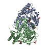
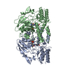
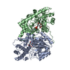
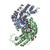
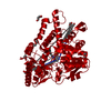
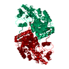
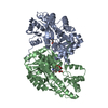
| Title | Crystal structure of Arabidopsis thaliana phosphoserine aminotransferase isoform 1 (AtPSAT1) in complex with Pyridoxamine-5'-phosphate (PMP) | ||||||
 Components Components | Phosphoserine aminotransferase 1, chloroplastic Phosphoserine transaminase Phosphoserine transaminase | ||||||
 Keywords Keywords | TRANSFERASE / serine biosynthesis / pyridoxal 5'-phosphate / PLP / transaminase / PSAT / pyridoxamine / PMP | ||||||
| Function / homology |  Function and homology informationphosphoserine transaminase / O-phospho-L-serine:2-oxoglutarate aminotransferase activity / L-serine biosynthetic process / plastid / chloroplast stroma / chloroplast / pyridoxal phosphate binding / protein homodimerization activity Function and homology informationphosphoserine transaminase / O-phospho-L-serine:2-oxoglutarate aminotransferase activity / L-serine biosynthetic process / plastid / chloroplast stroma / chloroplast / pyridoxal phosphate binding / protein homodimerization activitySimilarity search - Function | ||||||
| Biological species |  Arabidopsis thaliana (thale cress) Arabidopsis thaliana (thale cress) | ||||||
| Method | X-RAY DIFFRACTION / SYNCHROTRON / MOLECULAR REPLACEMENT / Resolution: 1.75 Å | ||||||
 Authors Authors | Sekula, B. / Ruszkowski, M. / Dauter, Z. | ||||||
| Funding support |  United States, 1items United States, 1items
| ||||||
 Citation Citation | Journal: Front Plant Sci / Year: 2018 Title: Structural Analysis of Phosphoserine Aminotransferase (Isoform 1) FromArabidopsis thaliana- the Enzyme Involved in the Phosphorylated Pathway of Serine Biosynthesis. Authors: Sekula, B. / Ruszkowski, M. / Dauter, Z. | ||||||
| History |
|
- Structure visualization
| Structure viewer | Molecule: MolmilJmol/JSmol |
|---|
- Downloads & links
-Download
| PDBx/mmCIF format | 6czy.cif.gz | 595.6 KB | Display | PDBx/mmCIF format |
|---|---|---|---|---|
| PDB format | pdb6czy.ent.gz | 491.6 KB | Display | PDB format |
| PDBx/mmJSON format | 6czy.json.gz | Tree view | PDBx/mmJSON format | |
| Others |  Other downloads Other downloads |
-Validation report
| Arichive directory | https://data.pdbj.org/pub/pdb/validation_reports/cz/6czyftp://data.pdbj.org/pub/pdb/validation_reports/cz/6czy | HTTPS FTP |
|---|
-Related structure data
| Related structure data | 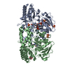 6czxC  6czzC  4xk1S S: Starting model for refinement C: citing same article ( |
|---|---|
| Similar structure data |


-Links
 PDBj
PDBj- Assembly
| Deposited unit | 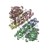
| ||||||||
|---|---|---|---|---|---|---|---|---|---|
| 1 | 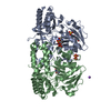
| ||||||||
| 2 | 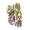
| ||||||||
| Unit cell |
|
-Components
-Protein , 1 types, 4 molecules ABCD
| #1: Protein | Phosphoserine transaminase / AtPSAT1 / Phosphohydroxythreonine aminotransferase Mass: 40276.176 Da / Num. of mol.: 4 Source method: isolated from a genetically manipulated source Source: (gene. exp.) Arabidopsis thaliana (thale cress) / Tissue: Leaves / Gene: PSAT1, At4g35630, F8D20.140 / Production host:  Escherichia coli (E. coli) / Strain (production host): BL21(DE3) / References: UniProt: Q96255, phosphoserine transaminase Escherichia coli (E. coli) / Strain (production host): BL21(DE3) / References: UniProt: Q96255, phosphoserine transaminase |
|---|
-Non-polymers , 6 types, 2046 molecules 


| #2: Chemical | ChemComp-PMP /  Mass: 248.173 Da / Num. of mol.: 4 / Source method: obtained synthetically / Formula: C8H13N2O5P Mass: 248.173 Da / Num. of mol.: 4 / Source method: obtained synthetically / Formula: C8H13N2O5P#3: Chemical | ChemComp-SO4 / Sulfate Mass: 96.063 Da / Num. of mol.: 7 / Source method: obtained synthetically / Formula: SO4 Mass: 96.063 Da / Num. of mol.: 7 / Source method: obtained synthetically / Formula: SO4#4: Chemical | Diethylene glycol Mass: 106.120 Da / Num. of mol.: 2 / Source method: obtained synthetically / Formula: C4H10O3 Mass: 106.120 Da / Num. of mol.: 2 / Source method: obtained synthetically / Formula: C4H10O3#5: Chemical | ChemComp-NA / |  Mass: 22.990 Da / Num. of mol.: 1 / Source method: obtained synthetically / Formula: Na Mass: 22.990 Da / Num. of mol.: 1 / Source method: obtained synthetically / Formula: Na#6: Chemical | ChemComp-MPD / ( | 2-Methyl-2,4-pentanediol Mass: 118.174 Da / Num. of mol.: 1 / Source method: obtained synthetically / Formula: C6H14O2 / Comment: precipitant*YM Mass: 118.174 Da / Num. of mol.: 1 / Source method: obtained synthetically / Formula: C6H14O2 / Comment: precipitant*YM#7: Water | ChemComp-HOH / | WaterMass: 18.015 Da / Num. of mol.: 2031 / Source method: isolated from a natural source / Formula: H2O |
|---|
-Experimental details
-Experiment
| Experiment | Method: X-RAY DIFFRACTION / Number of used crystals: 1 |
|---|
- Sample preparation
| Crystal | Density Matthews: 2.63 Å3/Da / Density % sol: 53.16 % |
|---|---|
| Crystal grow | Temperature: 292 K / Method: vapor diffusion, sitting drop / pH: 8.5 Details: 0.2 M lithium sulfate, 17% PEG 3350 and 0.1 M Tris at pH 8.5 |
-Data collection
| Diffraction | Mean temperature: 100 K |
|---|---|
| Diffraction source | Source: SYNCHROTRON / Site: APS / Beamline: 19-ID / Wavelength: 0.9792 Å |
| Detector | Type: DECTRIS PILATUS 6M / Detector: PIXEL / Date: Feb 8, 2018 |
| Radiation | Monochromator: Si (111) / Protocol: SINGLE WAVELENGTH / Monochromatic (M) / Laue (L): M / Scattering type: x-ray |
| Radiation wavelength | Wavelength: 0.9792 Å / Relative weight: 1 |
| Reflection | Resolution: 1.75→77.14 Å / Num. obs: 170626 / % possible obs: 99.4 % / Observed criterion σ(I): -3 / Redundancy: 7.3 % / Rmerge(I) obs: 0.102 / Net I/σ(I): 14.3 |
| Reflection shell | Resolution: 1.75→1.85 Å / Redundancy: 7.5 % / Rmerge(I) obs: 0.915 / Mean I/σ(I) obs: 2.14 / Num. unique obs: 26555 / % possible all: 96.6 |
- Processing
| Software |
| ||||||||||||||||||||||||||||||||||||||||||||||||||||||||||||||||||||||||||||||||||||||||||||||||||||||||||||||||||||||||||||||||||||||||||||||||||||||||||||||||||||||||||||||||||||||
|---|---|---|---|---|---|---|---|---|---|---|---|---|---|---|---|---|---|---|---|---|---|---|---|---|---|---|---|---|---|---|---|---|---|---|---|---|---|---|---|---|---|---|---|---|---|---|---|---|---|---|---|---|---|---|---|---|---|---|---|---|---|---|---|---|---|---|---|---|---|---|---|---|---|---|---|---|---|---|---|---|---|---|---|---|---|---|---|---|---|---|---|---|---|---|---|---|---|---|---|---|---|---|---|---|---|---|---|---|---|---|---|---|---|---|---|---|---|---|---|---|---|---|---|---|---|---|---|---|---|---|---|---|---|---|---|---|---|---|---|---|---|---|---|---|---|---|---|---|---|---|---|---|---|---|---|---|---|---|---|---|---|---|---|---|---|---|---|---|---|---|---|---|---|---|---|---|---|---|---|---|---|---|---|
| Refinement | Method to determine structure: MOLECULAR REPLACEMENT Starting model: 4XK1 Resolution: 1.75→77.14 Å / Cor.coef. Fo:Fc: 0.97 / Cor.coef. Fo:Fc free: 0.957 / SU B: 4.179 / SU ML: 0.07 / Cross valid method: THROUGHOUT / ESU R: 0.092 / ESU R Free: 0.093 / Details: HYDROGENS HAVE BEEN ADDED IN THE RIDING POSITIONS
| ||||||||||||||||||||||||||||||||||||||||||||||||||||||||||||||||||||||||||||||||||||||||||||||||||||||||||||||||||||||||||||||||||||||||||||||||||||||||||||||||||||||||||||||||||||||
| Solvent computation | Ion probe radii: 0.8 Å / Shrinkage radii: 0.8 Å / VDW probe radii: 1.2 Å | ||||||||||||||||||||||||||||||||||||||||||||||||||||||||||||||||||||||||||||||||||||||||||||||||||||||||||||||||||||||||||||||||||||||||||||||||||||||||||||||||||||||||||||||||||||||
| Displacement parameters | Biso mean: 22.936 Å2
| ||||||||||||||||||||||||||||||||||||||||||||||||||||||||||||||||||||||||||||||||||||||||||||||||||||||||||||||||||||||||||||||||||||||||||||||||||||||||||||||||||||||||||||||||||||||
| Refinement step | Cycle: 1 / Resolution: 1.75→77.14 Å
| ||||||||||||||||||||||||||||||||||||||||||||||||||||||||||||||||||||||||||||||||||||||||||||||||||||||||||||||||||||||||||||||||||||||||||||||||||||||||||||||||||||||||||||||||||||||
| Refine LS restraints |
|