 Movie
Movie Controller


+ Open data

- Basic information
| Entry | Database: PDB / ID: 1ugn | ||||||
|---|---|---|---|---|---|---|---|
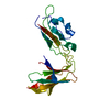
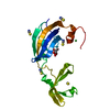
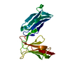
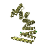
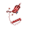
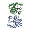
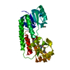
| Title | Crystal structure of LIR1.02, one of the alleles of LIR1 | ||||||
 Components Components | Leukocyte immunoglobulin-like receptor 1 | ||||||
 Keywords Keywords |  IMMUNE SYSTEM / immunoglobulin-like folds IMMUNE SYSTEM / immunoglobulin-like folds | ||||||
| Function / homology |  Function and homology information Function and homology informationHLA-A specific inhibitory MHC class I receptor activity / positive regulation of gamma-delta T cell activation involved in immune response / MHC class Ib protein complex binding / HLA-B specific inhibitory MHC class I receptor activity / inhibitory MHC class I receptor activity / negative regulation of dendritic cell differentiation / immune response-inhibiting cell surface receptor signaling pathway / MHC class Ib protein binding / Fc receptor mediated inhibitory signaling pathway / MHC class Ib receptor activity ...HLA-A specific inhibitory MHC class I receptor activity / positive regulation of gamma-delta T cell activation involved in immune response / MHC class Ib protein complex binding / HLA-B specific inhibitory MHC class I receptor activity / inhibitory MHC class I receptor activity / negative regulation of dendritic cell differentiation / immune response-inhibiting cell surface receptor signaling pathway / MHC class Ib protein binding / Fc receptor mediated inhibitory signaling pathway / MHC class Ib receptor activity / negative regulation of T cell mediated cytotoxicity / MHC class I receptor activity / negative regulation of CD8-positive, alpha-beta T cell activation / negative regulation of transforming growth factor beta production / negative regulation of alpha-beta T cell activation / negative regulation of cytokine production involved in immune response / negative regulation of serotonin secretion / dendritic cell differentiation / negative regulation of mononuclear cell proliferation / negative regulation of natural killer cell mediated cytotoxicity / negative regulation of T cell activation via T cell receptor contact with antigen bound to MHC molecule on antigen presenting cell / protein phosphatase 1 binding / negative regulation of osteoclast development / negative regulation of interleukin-12 production / negative regulation of endocytosis / negative regulation of interferon-beta production / negative regulation of dendritic cell apoptotic process / positive regulation of macrophage cytokine production / negative regulation of interleukin-10 production / T cell proliferation involved in immune response / MHC class I protein binding / negative regulation of calcium ion transport / negative regulation of type II interferon production / negative regulation of tumor necrosis factor production / negative regulation of cell cycle / negative regulation of T cell proliferation / positive regulation of defense response to virus by host / SH2 domain binding / response to virus / receptor internalization / cytokine-mediated signaling pathway / Immunoregulatory interactions between a Lymphoid and a non-Lymphoid cell / positive regulation of type II interferon production / defense response to virus / adaptive immune response / cellular response to lipopolysaccharide / positive regulation of apoptotic process / external side of plasma membrane / positive regulation of gene expression / signal transduction / protein homodimerization activity / positive regulation of transcription by RNA polymerase II / extracellular region / plasma membrane / cytoplasmSimilarity search - Function | ||||||
| Biological species |  Homo sapiens (human) Homo sapiens (human) | ||||||
| Method | X-RAY DIFFRACTION / SYNCHROTRON / MOLECULAR REPLACEMENT / Resolution: 1.8 Å | ||||||
 Authors Authors | Shiroishi, M. / Rasubala, L. / Kuroki, K. / Amano, K. / Tsuchiya, N. / Tokunaga, K. / Kohda, D. / Maenaka, K. | ||||||
 Citation Citation | Journal: Hum.Mol.Genet. / Year: 2005 Title: Extensive polymorphisms of LILRB1 (ILT2, LIR1) and their association with HLA-DRB1 shared epitope negative rheumatoid arthritis. Authors: Kuroki, K. / Tsuchiya, N. / Shiroishi, M. / Rasubala, L. / Yamashita, Y. / Matsuta, K. / Fukazawa, T. / Kusaoi, M. / Murakami, Y. / Takiguchi, M. / Juji, T. / Hashimoto, H. / Kohda, D. / ...Authors: Kuroki, K. / Tsuchiya, N. / Shiroishi, M. / Rasubala, L. / Yamashita, Y. / Matsuta, K. / Fukazawa, T. / Kusaoi, M. / Murakami, Y. / Takiguchi, M. / Juji, T. / Hashimoto, H. / Kohda, D. / Maenaka, K. / Tokunaga, K. | ||||||
| History |
|
- Structure visualization
| Structure viewer | Molecule: MolmilJmol/JSmol |
|---|
- Downloads & links
-Download
| PDBx/mmCIF format | 1ugn.cif.gz | 53.6 KB | Display | PDBx/mmCIF format |
|---|---|---|---|---|
| PDB format | pdb1ugn.ent.gz | 37.5 KB | Display | PDB format |
| PDBx/mmJSON format | 1ugn.json.gz | Tree view | PDBx/mmJSON format | |
| Others |  Other downloads Other downloads |
-Validation report
| Arichive directory | https://data.pdbj.org/pub/pdb/validation_reports/ug/1ugnftp://data.pdbj.org/pub/pdb/validation_reports/ug/1ugn | HTTPS FTP |
|---|
-Related structure data
| Related structure data |  1ufuS S: Starting model for refinement |
|---|---|
| Similar structure data |


-Links
 PDBj
PDBj


- Assembly
| Deposited unit | 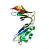
| ||||||||
|---|---|---|---|---|---|---|---|---|---|
| 1 |
| ||||||||
| Unit cell |
|
-Components
| #1: Protein | Mass: 22085.723 Da / Num. of mol.: 1 / Fragment: Ligand binding domain (domain1 and 2) / Mutation: A70T Source method: isolated from a genetically manipulated source Source: (gene. exp.) Homo sapiens (human) / Plasmid: pGMT7 / Species (production host): Escherichia coli / Production host:  Escherichia coli BL21(DE3) (bacteria) / Strain (production host): BL21(DE3) / References: UniProt: Q8NHL6 Escherichia coli BL21(DE3) (bacteria) / Strain (production host): BL21(DE3) / References: UniProt: Q8NHL6 |
|---|---|
| #2: Water | ChemComp-HOH / Water Mass: 18.015 Da / Num. of mol.: 175 / Source method: isolated from a natural source / Formula: H2O Mass: 18.015 Da / Num. of mol.: 175 / Source method: isolated from a natural source / Formula: H2O |
-Experimental details
-Experiment
| Experiment | Method: X-RAY DIFFRACTION / Number of used crystals: 1 |
|---|
- Sample preparation
| Crystal | Density Matthews: 2.18 Å3/Da / Density % sol: 43.05 % |
|---|---|
| Crystal grow | Temperature: 298 K / Method: vapor diffusion, sitting drop / pH: 8.5 Details: 0.1M Tris chloride, 0.7M potassium sodium tartrate, pH 8.5, VAPOR DIFFUSION, SITTING DROP, temperature 298K |
-Data collection
| Diffraction | Mean temperature: 100 K |
|---|---|
| Diffraction source | Source: SYNCHROTRON / Site: SPring-8  / Beamline: BL41XU / Wavelength: 0.984 Å / Beamline: BL41XU / Wavelength: 0.984 Å |
| Detector | Type: MARRESEARCH / Detector: CCD |
| Radiation | Protocol: SINGLE WAVELENGTH / Monochromatic (M) / Laue (L): M / Scattering type: x-ray |
| Radiation wavelength | Wavelength: 0.984 Å / Relative weight: 1 |
| Reflection | Resolution: 1.8→50 Å / Num. obs: 18378 / % possible obs: 95.9 % / Observed criterion σ(I): 1 / Redundancy: 5.8 % / Biso Wilson estimate: 17.8 Å2 / Rmerge(I) obs: 0.097 / Net I/σ(I): 6.9 |
| Reflection shell | Resolution: 1.8→1.86 Å / Redundancy: 3.6 % / Rmerge(I) obs: 0.465 / Mean I/σ(I) obs: 2.03 / Num. unique all: 1539 / % possible all: 83.1 |
- Processing
| Software |
| ||||||||||||||||||||
|---|---|---|---|---|---|---|---|---|---|---|---|---|---|---|---|---|---|---|---|---|---|
| Refinement | Method to determine structure: MOLECULAR REPLACEMENT Starting model: PDB entry 1UFU Resolution: 1.8→8 Å / Isotropic thermal model: isotropic / σ(F): 0 / Stereochemistry target values: Engh & Huber
| ||||||||||||||||||||
| Displacement parameters | Biso mean: 21.6 Å2 | ||||||||||||||||||||
| Refine analyze |
| ||||||||||||||||||||
| Refinement step | Cycle: LAST / Resolution: 1.8→8 Å
| ||||||||||||||||||||
| Refine LS restraints |
| ||||||||||||||||||||
| LS refinement shell | Resolution: 1.8→1.91 Å / Rfactor Rfree error: 0.027
|
