 Movie
Movie Controller


+ Open data

- Basic information
| Entry | Database: PDB / ID: 3cgm | ||||||
|---|---|---|---|---|---|---|---|
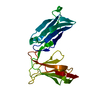
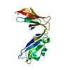
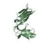
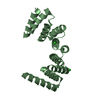
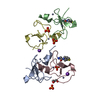
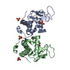
| Title | Crystal structure of thermophilic SlyD | ||||||
 Components Components | Peptidyl-prolyl cis-trans isomerase Prolyl isomerase Prolyl isomerase | ||||||
 Keywords Keywords | ISOMERASE / Prolyl cis trans isomerase / chaperone function / two domain protein / Rotamase | ||||||
| Function / homology |  Function and homology informationpeptidylprolyl isomerase / peptidyl-prolyl cis-trans isomerase activity / protein refolding / metal ion binding Function and homology informationpeptidylprolyl isomerase / peptidyl-prolyl cis-trans isomerase activity / protein refolding / metal ion bindingSimilarity search - Function | ||||||
| Biological species |   Thermus thermophilus (bacteria) Thermus thermophilus (bacteria) | ||||||
| Method | X-RAY DIFFRACTION / SYNCHROTRON / molecular replacement / Resolution: 2.41 Å | ||||||
 Authors Authors | Loew, C. / Neumann, P. / Stubbs, M.T. / Balbach, J. | ||||||
 Citation Citation | Journal: J.Mol.Biol. / Year: 2010 Title: Crystal Structure Determination and Functional Characterization of the Metallochaperone SlyD from Thermus thermophilus Authors: Loew, C. / Neumann, P. / Tidow, H. / Weininger, U. / Haupt, C. / Friedrich-Epler, B. / Scholz, C. / Stubbs, M.T. / Balbach, J. | ||||||
| History |
|
- Structure visualization
| Structure viewer | Molecule: MolmilJmol/JSmol |
|---|
- Downloads & links
-Download
| PDBx/mmCIF format | 3cgm.cif.gz | 80.6 KB | Display | PDBx/mmCIF format |
|---|---|---|---|---|
| PDB format | pdb3cgm.ent.gz | 60.7 KB | Display | PDB format |
| PDBx/mmJSON format | 3cgm.json.gz | Tree view | PDBx/mmJSON format | |
| Others |  Other downloads Other downloads |
-Validation report
| Arichive directory | https://data.pdbj.org/pub/pdb/validation_reports/cg/3cgmftp://data.pdbj.org/pub/pdb/validation_reports/cg/3cgm | HTTPS FTP |
|---|
-Related structure data
| Related structure data | 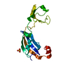 3cgnC  3luoC  1d6oS C: citing same article ( S: Starting model for refinement |
|---|---|
| Similar structure data |


-Links
 PDBj
PDBj


- Assembly
| Deposited unit | 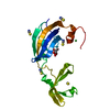
| ||||||||
|---|---|---|---|---|---|---|---|---|---|
| 1 |
| ||||||||
| 2 | x 6
| ||||||||
| Unit cell |
|
-Components
| #1: Protein | Prolyl isomerase / SlyD Mass: 17400.234 Da / Num. of mol.: 1 Source method: isolated from a genetically manipulated source Source: (gene. exp.) Thermus thermophilus (bacteria) / Strain: HB8 / Plasmid: pet11a / Production host: Escherichia coli (E. coli) / Strain (production host): BL21 / References: UniProt: Q5SLE7, peptidylprolyl isomerase | ||||||
|---|---|---|---|---|---|---|---|
| #2: Chemical | ChemComp-SCN / Thiocyanate  Mass: 58.082 Da / Num. of mol.: 6 / Source method: obtained synthetically / Formula: CNS Mass: 58.082 Da / Num. of mol.: 6 / Source method: obtained synthetically / Formula: CNS#3: Chemical | ChemComp-NI / | Nickel  Mass: 58.693 Da / Num. of mol.: 1 / Source method: obtained synthetically / Formula: Ni Mass: 58.693 Da / Num. of mol.: 1 / Source method: obtained synthetically / Formula: Ni#4: Chemical | Glycerol  Mass: 92.094 Da / Num. of mol.: 2 / Source method: obtained synthetically / Formula: C3H8O3 Mass: 92.094 Da / Num. of mol.: 2 / Source method: obtained synthetically / Formula: C3H8O3#5: Water | ChemComp-HOH / | Water Mass: 18.015 Da / Num. of mol.: 98 / Source method: isolated from a natural source / Formula: H2O Mass: 18.015 Da / Num. of mol.: 98 / Source method: isolated from a natural source / Formula: H2O |
-Experimental details
-Experiment
| Experiment | Method: X-RAY DIFFRACTION / Number of used crystals: 1 |
|---|
- Sample preparation
| Crystal | Density Matthews: 4.69 Å3/Da / Density % sol: 73.78 % |
|---|---|
| Crystal grow | Temperature: 286 K / Method: vapor diffusion, hanging drop / pH: 7.5 Details: 60mg/ml protein, 1:1 mixing, 2M Ammonium sulfate, 2% PEG 400, 1% Thiocyanate, 100mM HEPES, pH7.5, VAPOR DIFFUSION, HANGING DROP, temperature 286K |
-Data collection
| Diffraction | Mean temperature: 180 K |
|---|---|
| Diffraction source | Source: SYNCHROTRON / Site: BESSY  / Beamline: 14.1 / Wavelength: 0.9184 Å / Beamline: 14.1 / Wavelength: 0.9184 Å |
| Detector | Type: MARMOSAIC 225 mm CCD / Detector: CCD / Date: Jul 7, 2007 / Details: mirrors |
| Radiation | Monochromator: GRAPHITE / Protocol: SINGLE WAVELENGTH / Monochromatic (M) / Laue (L): M / Scattering type: x-ray |
| Radiation wavelength | Wavelength: 0.9184 Å / Relative weight: 1 |
| Reflection | Resolution: 2.41→30 Å / Num. all: 13374 / Num. obs: 12921 / % possible obs: 96.6 % / Observed criterion σ(F): 0 / Observed criterion σ(I): -3 / Redundancy: 13.1 % / Biso Wilson estimate: 43.405 Å2 / Rmerge(I) obs: 0.093 / Rsym value: 0.107 / Net I/σ(I): 21.98 |
| Reflection shell | Resolution: 2.41→2.55 Å / Redundancy: 11.32 % / Rmerge(I) obs: 0.582 / Mean I/σ(I) obs: 4.4 / Num. measured obs: 21342 / Num. unique all: 1885 / Num. unique obs: 1885 / Rsym value: 0.45 / % possible all: 92.9 |
- Processing
| Software |
| |||||||||||||||||||||||||||||||||||||||||||||||||||||||||||||||||||||||||||
|---|---|---|---|---|---|---|---|---|---|---|---|---|---|---|---|---|---|---|---|---|---|---|---|---|---|---|---|---|---|---|---|---|---|---|---|---|---|---|---|---|---|---|---|---|---|---|---|---|---|---|---|---|---|---|---|---|---|---|---|---|---|---|---|---|---|---|---|---|---|---|---|---|---|---|---|---|
| Refinement | Method to determine structure: molecular replacement Starting model: PDB ENTRY 1D6O Resolution: 2.41→28.27 Å / Cor.coef. Fo:Fc: 0.943 / Cor.coef. Fo:Fc free: 0.924 / SU B: 10.394 / SU ML: 0.129 / Isotropic thermal model: Isotropic/TLS / Cross valid method: THROUGHOUT / σ(F): 0 / σ(I): -3 / ESU R: 0.214 / ESU R Free: 0.19 / Stereochemistry target values: MAXIMUM LIKELIHOOD
| |||||||||||||||||||||||||||||||||||||||||||||||||||||||||||||||||||||||||||
| Solvent computation | Ion probe radii: 0.8 Å / Shrinkage radii: 0.8 Å / VDW probe radii: 1.2 Å / Solvent model: BABINET MODEL WITH MASK | |||||||||||||||||||||||||||||||||||||||||||||||||||||||||||||||||||||||||||
| Displacement parameters | Biso mean: 44.556 Å2
| |||||||||||||||||||||||||||||||||||||||||||||||||||||||||||||||||||||||||||
| Refine analyze |
| |||||||||||||||||||||||||||||||||||||||||||||||||||||||||||||||||||||||||||
| Refinement step | Cycle: LAST / Resolution: 2.41→28.27 Å
| |||||||||||||||||||||||||||||||||||||||||||||||||||||||||||||||||||||||||||
| Refine LS restraints |
| |||||||||||||||||||||||||||||||||||||||||||||||||||||||||||||||||||||||||||
| LS refinement shell | Resolution: 2.409→2.471 Å / Rfactor Rfree error: 0 / Total num. of bins used: 20
| |||||||||||||||||||||||||||||||||||||||||||||||||||||||||||||||||||||||||||
| Refinement TLS params. | Method: refined / Refine-ID: X-RAY DIFFRACTION
| |||||||||||||||||||||||||||||||||||||||||||||||||||||||||||||||||||||||||||
| Refinement TLS group |
|
