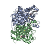
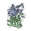
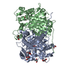
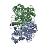
 Movie
Movie Controller

[English] 日本語
 Yorodumi
- PDB-1u6e: 1.85 Angstrom Crystal Structure of the C112A Mutant of Mycobacter... -
+ Open data

- Basic information
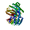
| Entry | Database: PDB / ID: 1u6e | ||||||
|---|---|---|---|---|---|---|---|
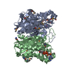
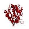
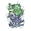
| Title | 1.85 Angstrom Crystal Structure of the C112A Mutant of Mycobacterium Tuberculosis Beta-Ketoacyl-Acyl Carrier Protein Synthase III (FabH) | ||||||
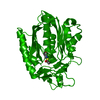
 Components Components | 3-oxoacyl-[acyl-carrier-protein] synthase III | ||||||
 Keywords Keywords |  TRANSFERASE / 3-oxoacyl-[acyl-carrier-protein] synthase III TRANSFERASE / 3-oxoacyl-[acyl-carrier-protein] synthase III | ||||||
| Function / homology |  Function and homology information Function and homology informationmycobacterial beta-ketoacyl-[acyl carrier protein] synthase III / beta-ketodecanoyl-[acyl-carrier-protein] synthase activity / long-chain fatty-acyl-CoA metabolic process / beta-ketoacyl-[acyl-carrier-protein] synthase III / beta-ketoacyl-acyl-carrier-protein synthase III activity / fatty-acyl-CoA binding / secondary metabolite biosynthetic process / fatty acid elongation / lipid biosynthetic process / 3-oxoacyl-[acyl-carrier-protein] synthase activity ...mycobacterial beta-ketoacyl-[acyl carrier protein] synthase III / beta-ketodecanoyl-[acyl-carrier-protein] synthase activity / long-chain fatty-acyl-CoA metabolic process / beta-ketoacyl-[acyl-carrier-protein] synthase III / beta-ketoacyl-acyl-carrier-protein synthase III activity / fatty-acyl-CoA binding / secondary metabolite biosynthetic process / fatty acid elongation / lipid biosynthetic process / 3-oxoacyl-[acyl-carrier-protein] synthase activity / fatty acid biosynthetic process / cytoplasmSimilarity search - Function | ||||||
| Biological species |   Mycobacterium tuberculosis (bacteria) Mycobacterium tuberculosis (bacteria) | ||||||
| Method | X-RAY DIFFRACTION / MOLECULAR REPLACEMENT / Resolution: 1.85 Å | ||||||
 Authors Authors | Mussayev, F. / Sachedeva, S. / Scarsdale, J.N. / Reynolds, K.A. / Wright, H.T. | ||||||
 Citation Citation | Journal: J.Mol.Biol. / Year: 2005 Title: Crystal structure of a substrate complex of Mycobacterium tuberculosis beta-ketoacyl-acyl carrier protein synthase III (FabH) with lauroyl-coenzyme A. Authors: Musayev, F. / Sachdeva, S. / Scarsdale, J.N. / Reynolds, K.A. / Wright, H.T. #1: Journal: J.Biol.Chem. / Year: 2001Title: Crystal Structure of the Mycobacterium Tuberculosis beta- ketoacyl-acyl carrier protein synthase III Authors: Scarsdale, J.N. / Kazanina, G. / He, X. / Reynolds, K.A. / Wright, H.T. | ||||||
| History |
|
- Structure visualization
| Structure viewer | Molecule: MolmilJmol/JSmol |
|---|
- Downloads & links
-Download
| PDBx/mmCIF format | 1u6e.cif.gz | 145.9 KB | Display | PDBx/mmCIF format |
|---|---|---|---|---|
| PDB format | pdb1u6e.ent.gz | 112 KB | Display | PDB format |
| PDBx/mmJSON format | 1u6e.json.gz | Tree view | PDBx/mmJSON format | |
| Others |  Other downloads Other downloads |
-Validation report
| Arichive directory | https://data.pdbj.org/pub/pdb/validation_reports/u6/1u6eftp://data.pdbj.org/pub/pdb/validation_reports/u6/1u6e | HTTPS FTP |
|---|
-Related structure data
| Related structure data | 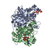 1u6sC  1hzpS S: Starting model for refinement C: citing same article ( |
|---|---|
| Similar structure data |


-Links
 PDBj
PDBj

- Assembly
| Deposited unit | 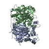
| |||||||||||||||||||||||||||||||||||||||||||||||||||||||||||||||||||||||||||||||||||||||||||||||||||||||||||||||||||||||||||||||||||||||||||||||||||||||||||||||||||||||||||||||||||||||||||||||||||
|---|---|---|---|---|---|---|---|---|---|---|---|---|---|---|---|---|---|---|---|---|---|---|---|---|---|---|---|---|---|---|---|---|---|---|---|---|---|---|---|---|---|---|---|---|---|---|---|---|---|---|---|---|---|---|---|---|---|---|---|---|---|---|---|---|---|---|---|---|---|---|---|---|---|---|---|---|---|---|---|---|---|---|---|---|---|---|---|---|---|---|---|---|---|---|---|---|---|---|---|---|---|---|---|---|---|---|---|---|---|---|---|---|---|---|---|---|---|---|---|---|---|---|---|---|---|---|---|---|---|---|---|---|---|---|---|---|---|---|---|---|---|---|---|---|---|---|---|---|---|---|---|---|---|---|---|---|---|---|---|---|---|---|---|---|---|---|---|---|---|---|---|---|---|---|---|---|---|---|---|---|---|---|---|---|---|---|---|---|---|---|---|---|---|---|---|---|
| 1 |
| |||||||||||||||||||||||||||||||||||||||||||||||||||||||||||||||||||||||||||||||||||||||||||||||||||||||||||||||||||||||||||||||||||||||||||||||||||||||||||||||||||||||||||||||||||||||||||||||||||
| Unit cell |
| |||||||||||||||||||||||||||||||||||||||||||||||||||||||||||||||||||||||||||||||||||||||||||||||||||||||||||||||||||||||||||||||||||||||||||||||||||||||||||||||||||||||||||||||||||||||||||||||||||
| Noncrystallographic symmetry (NCS) | NCS domain:
NCS domain segments: Ens-ID: 1 / Refine code: 4
|