 Movie
Movie Controller

[English] 日本語
 Yorodumi
- PDB-1hzp: Crystal Structure of the Myobacterium Tuberculosis Beta-Ketoacyl-... -
+ Open data

- Basic information
| Entry | Database: PDB / ID: 1hzp | ||||||
|---|---|---|---|---|---|---|---|
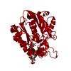
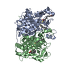
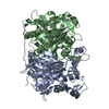
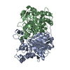
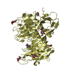
| Title | Crystal Structure of the Myobacterium Tuberculosis Beta-Ketoacyl-Acyl Carrier Protein Synthase III | ||||||
 Components Components | 3-OXOACYL-[ACYL-CARRIER-PROTEIN] SYNTHASE III | ||||||
 Keywords Keywords |  TRANSFERASE / fatty acid biosynthesis / myobacterium tuberculosis / structural basis for substrate specificity / Structural Genomics / PSI / Protein Structure Initiative / TB Structural Genomics Consortium / TBSGC TRANSFERASE / fatty acid biosynthesis / myobacterium tuberculosis / structural basis for substrate specificity / Structural Genomics / PSI / Protein Structure Initiative / TB Structural Genomics Consortium / TBSGC | ||||||
| Function / homology |  Function and homology information Function and homology informationmycobacterial beta-ketoacyl-[acyl carrier protein] synthase III / beta-ketodecanoyl-[acyl-carrier-protein] synthase activity / long-chain fatty-acyl-CoA metabolic process / beta-ketoacyl-[acyl-carrier-protein] synthase III / beta-ketoacyl-acyl-carrier-protein synthase III activity / fatty-acyl-CoA binding / fatty acid elongation / lipid biosynthetic process / 3-oxoacyl-[acyl-carrier-protein] synthase activity / fatty acid biosynthetic process / cytoplasmSimilarity search - Function | ||||||
| Biological species |   Mycobacterium tuberculosis (bacteria) Mycobacterium tuberculosis (bacteria) | ||||||
| Method | X-RAY DIFFRACTION / MOLECULAR REPLACEMENT / Resolution: 2.1 Å | ||||||
 Authors Authors | Scarsdale, J.N. / Kazanina, G. / He, X. / Reynolds, K.A. / Wright, H.T. / TB Structural Genomics Consortium (TBSGC) | ||||||
 Citation Citation | Journal: J.Biol.Chem. / Year: 2001 Title: Crystal structure of the Mycobacterium tuberculosis beta-ketoacyl-acyl carrier protein synthase III Authors: Scarsdale, J.N. / Kazanina, G. / He, X. / Reynolds, K.A. / Wright, H.T. | ||||||
| History |
|
- Structure visualization
| Structure viewer | Molecule: MolmilJmol/JSmol |
|---|
- Downloads & links
-Download
| PDBx/mmCIF format | 1hzp.cif.gz | 137.8 KB | Display | PDBx/mmCIF format |
|---|---|---|---|---|
| PDB format | pdb1hzp.ent.gz | 106 KB | Display | PDB format |
| PDBx/mmJSON format | 1hzp.json.gz | Tree view | PDBx/mmJSON format | |
| Others |  Other downloads Other downloads |
-Validation report
| Arichive directory | https://data.pdbj.org/pub/pdb/validation_reports/hz/1hzpftp://data.pdbj.org/pub/pdb/validation_reports/hz/1hzp | HTTPS FTP |
|---|
-Related structure data
| Related structure data | 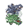 1eblS S: Starting model for refinement |
|---|---|
| Similar structure data | |
| Other databases |


-Links
 PDBj
PDBj


- Assembly
| Deposited unit | 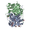
| ||||||||
|---|---|---|---|---|---|---|---|---|---|
| 1 |
| ||||||||
| Unit cell |
|
-Components
| #1: Protein | Mass: 34903.391 Da / Num. of mol.: 2 Source method: isolated from a genetically manipulated source Source: (gene. exp.) Mycobacterium tuberculosis (bacteria) / Plasmid: PET15B / Production host: Escherichia coli (E. coli) / Strain (production host): BL21(DE3)PLYSSReferences: UniProt: P0A574, UniProt: P9WNG3*PLUS, beta-ketoacyl-[acyl-carrier-protein] synthase I #2: Chemical | ChemComp-DAO / | Lauric acid  Mass: 200.318 Da / Num. of mol.: 1 / Source method: obtained synthetically / Formula: C12H24O2 Mass: 200.318 Da / Num. of mol.: 1 / Source method: obtained synthetically / Formula: C12H24O2#3: Chemical | ChemComp-GOL / Glycerol  Mass: 92.094 Da / Num. of mol.: 7 / Source method: obtained synthetically / Formula: C3H8O3 Mass: 92.094 Da / Num. of mol.: 7 / Source method: obtained synthetically / Formula: C3H8O3#4: Water | ChemComp-HOH / | Water Mass: 18.015 Da / Num. of mol.: 258 / Source method: isolated from a natural source / Formula: H2O Mass: 18.015 Da / Num. of mol.: 258 / Source method: isolated from a natural source / Formula: H2O |
|---|
-Experimental details
-Experiment
| Experiment | Method: X-RAY DIFFRACTION / Number of used crystals: 1 |
|---|
- Sample preparation
| Crystal | Density Matthews: 2.21 Å3/Da / Density % sol: 43.9 % | ||||||||||||||||||||||||||||||||||||||||||||||||||||||||||||||||||
|---|---|---|---|---|---|---|---|---|---|---|---|---|---|---|---|---|---|---|---|---|---|---|---|---|---|---|---|---|---|---|---|---|---|---|---|---|---|---|---|---|---|---|---|---|---|---|---|---|---|---|---|---|---|---|---|---|---|---|---|---|---|---|---|---|---|---|---|
| Crystal grow | Temperature: 298 K / Method: vapor diffusion / pH: 8 Details: PEG4000, Tris/HCl. 300mM NaCl, glycerol, 10mM CaCl2, pH 8.0, VAPOR DIFFUSION, temperature 298K | ||||||||||||||||||||||||||||||||||||||||||||||||||||||||||||||||||
| Crystal grow | *PLUS | ||||||||||||||||||||||||||||||||||||||||||||||||||||||||||||||||||
| Components of the solutions | *PLUS
|
-Data collection
| Diffraction | Mean temperature: 113 K |
|---|---|
| Diffraction source | Source: ROTATING ANODE / Type: RIGAKU / Wavelength: 1.5418 Å |
| Detector | Type: RIGAKU RAXIS II / Detector: IMAGE PLATE / Details: Osmic Confocal optics |
| Radiation | Monochromator: Osmic Confocal optics / Protocol: SINGLE WAVELENGTH / Monochromatic (M) / Laue (L): M / Scattering type: x-ray |
| Radiation wavelength | Wavelength: 1.5418 Å / Relative weight: 1 |
| Reflection | Resolution: 2.1→99 Å / Num. all: 37716 / Num. obs: 37503 / % possible obs: 99.4 % / Observed criterion σ(I): -3 / Redundancy: 3 % / Biso Wilson estimate: 26.4 Å2 / Rmerge(I) obs: 0.119 / Net I/σ(I): 8.3 |
| Reflection shell | Resolution: 2.1→2.14 Å / Redundancy: 2.7 % / Rmerge(I) obs: 0.688 / Mean I/σ(I) obs: 1.8 / Num. unique all: 1860 / % possible all: 99 |
| Reflection shell | *PLUS % possible obs: 82 % |
- Processing
| Software |
| ||||||||||||||||||||||||||||||||||||||||||||||||||||||||||||||||||||||||||||||||
|---|---|---|---|---|---|---|---|---|---|---|---|---|---|---|---|---|---|---|---|---|---|---|---|---|---|---|---|---|---|---|---|---|---|---|---|---|---|---|---|---|---|---|---|---|---|---|---|---|---|---|---|---|---|---|---|---|---|---|---|---|---|---|---|---|---|---|---|---|---|---|---|---|---|---|---|---|---|---|---|---|---|
| Refinement | Method to determine structure: MOLECULAR REPLACEMENT Starting model: poly alanine chain based on residues 1-317 from E Coli FabH (RCSB entry 1EBL) Resolution: 2.1→30.38 Å / Rfactor Rfree error: 0.004 / Data cutoff high absF: 797513.75 / Data cutoff low absF: 0 / Isotropic thermal model: RESTRAINED / Cross valid method: THROUGHOUT / σ(F): 2 / Stereochemistry target values: Engh & Huber
| ||||||||||||||||||||||||||||||||||||||||||||||||||||||||||||||||||||||||||||||||
| Solvent computation | Solvent model: FLAT MODEL / Bsol: 63.46 Å2 / ksol: 0.366 e/Å3 | ||||||||||||||||||||||||||||||||||||||||||||||||||||||||||||||||||||||||||||||||
| Displacement parameters | Biso mean: 31.7 Å2
| ||||||||||||||||||||||||||||||||||||||||||||||||||||||||||||||||||||||||||||||||
| Refine analyze |
| ||||||||||||||||||||||||||||||||||||||||||||||||||||||||||||||||||||||||||||||||
| Refinement step | Cycle: LAST / Resolution: 2.1→30.38 Å
| ||||||||||||||||||||||||||||||||||||||||||||||||||||||||||||||||||||||||||||||||
| Refine LS restraints |
| ||||||||||||||||||||||||||||||||||||||||||||||||||||||||||||||||||||||||||||||||
| LS refinement shell | Resolution: 2.1→2.23 Å / Rfactor Rfree error: 0.013 / Total num. of bins used: 6
| ||||||||||||||||||||||||||||||||||||||||||||||||||||||||||||||||||||||||||||||||
| Xplor file |
| ||||||||||||||||||||||||||||||||||||||||||||||||||||||||||||||||||||||||||||||||
| Software | *PLUS Name: CNS / Version: 1 / Classification: refinement | ||||||||||||||||||||||||||||||||||||||||||||||||||||||||||||||||||||||||||||||||
| Refinement | *PLUS σ(F): 2 / % reflection Rfree: 10 % | ||||||||||||||||||||||||||||||||||||||||||||||||||||||||||||||||||||||||||||||||
| Solvent computation | *PLUS | ||||||||||||||||||||||||||||||||||||||||||||||||||||||||||||||||||||||||||||||||
| Displacement parameters | *PLUS Biso mean: 31.7 Å2 | ||||||||||||||||||||||||||||||||||||||||||||||||||||||||||||||||||||||||||||||||
| Refine LS restraints | *PLUS
| ||||||||||||||||||||||||||||||||||||||||||||||||||||||||||||||||||||||||||||||||
| LS refinement shell | *PLUS Rfactor Rfree: 0.319 / % reflection Rfree: 10 % / Rfactor Rwork: 0.281 / Rfactor obs: 0.281 |
