 ムービー
ムービー コントローラー


+ データを開く

- 基本情報
基本情報
| 登録情報 | データベース: PDB / ID: 1u2c | ||||||
|---|---|---|---|---|---|---|---|
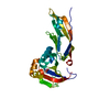
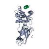
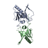
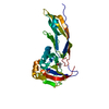
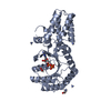
| タイトル | Crystal Structure of a-dystroglycan | ||||||
 要素 要素 | Dystroglycan | ||||||
 キーワード キーワード | PROTEIN BINDING (タンパク質) / IG-like domain / S6 like fold | ||||||
| 機能・相同性 |  機能・相同性情報O-結合型グリコシル化 / dystroglycan complex / nerve maturation / muscle attachment / positive regulation of basement membrane assembly involved in embryonic body morphogenesis / retrograde trans-synaptic signaling by trans-synaptic protein complex / Regulation of expression of SLITs and ROBOs / contractile ring / regulation of gastrulation / microtubule anchoring ...O-結合型グリコシル化 / dystroglycan complex / nerve maturation / muscle attachment / positive regulation of basement membrane assembly involved in embryonic body morphogenesis / retrograde trans-synaptic signaling by trans-synaptic protein complex / Regulation of expression of SLITs and ROBOs / contractile ring / regulation of gastrulation / microtubule anchoring / calcium-dependent cell-matrix adhesion / morphogenesis of an epithelial sheet / dystrophin-associated glycoprotein complex / laminin-1 binding / response to denervation involved in regulation of muscle adaptation / basement membrane organization / positive regulation of myelination / regulation of epithelial to mesenchymal transition / skeletal muscle tissue regeneration / dystroglycan binding / cellular response to cholesterol / nerve development / vinculin binding / photoreceptor ribbon synapse / myelination in peripheral nervous system / branching involved in salivary gland morphogenesis / ランヴィエの絞輪 / costamere / angiogenesis involved in wound healing / commissural neuron axon guidance / response to muscle activity / regulation of neurotransmitter receptor localization to postsynaptic specialization membrane / postsynaptic cytosol / axon regeneration / positive regulation of cell-matrix adhesion / structural constituent of muscle / negative regulation of MAPK cascade / epithelial tube branching involved in lung morphogenesis / positive regulation of oligodendrocyte differentiation / regulation of synapse organization / alpha-actinin binding / plasma membrane raft / membrane protein ectodomain proteolysis / cellular response to organic cyclic compound / 基底膜 / GABA-ergic synapse / negative regulation of phosphatidylinositol 3-kinase/protein kinase B signal transduction / laminin binding / Schwann cell development / heart morphogenesis / SH2 domain binding / tubulin binding / nuclear periphery / negative regulation of cell migration / filopodium / morphogenesis of an epithelium / 軸索誘導 / 接着結合 / regulation of synaptic plasticity / 筋鞘 / response to peptide hormone / cellular response to mechanical stimulus / cell-cell junction / protein transport / virus receptor activity / lamellipodium / actin binding / heart development / postsynapse / basolateral plasma membrane / postsynaptic membrane / 細胞骨格 / 脂質ラフト / external side of plasma membrane / intracellular membrane-bounded organelle / focal adhesion / glutamatergic synapse / calcium ion binding / protein-containing complex binding / 細胞膜 / extracellular space / extracellular region / 核質 / 生体膜 / 細胞膜 / 細胞質 機能・相同性情報O-結合型グリコシル化 / dystroglycan complex / nerve maturation / muscle attachment / positive regulation of basement membrane assembly involved in embryonic body morphogenesis / retrograde trans-synaptic signaling by trans-synaptic protein complex / Regulation of expression of SLITs and ROBOs / contractile ring / regulation of gastrulation / microtubule anchoring ...O-結合型グリコシル化 / dystroglycan complex / nerve maturation / muscle attachment / positive regulation of basement membrane assembly involved in embryonic body morphogenesis / retrograde trans-synaptic signaling by trans-synaptic protein complex / Regulation of expression of SLITs and ROBOs / contractile ring / regulation of gastrulation / microtubule anchoring / calcium-dependent cell-matrix adhesion / morphogenesis of an epithelial sheet / dystrophin-associated glycoprotein complex / laminin-1 binding / response to denervation involved in regulation of muscle adaptation / basement membrane organization / positive regulation of myelination / regulation of epithelial to mesenchymal transition / skeletal muscle tissue regeneration / dystroglycan binding / cellular response to cholesterol / nerve development / vinculin binding / photoreceptor ribbon synapse / myelination in peripheral nervous system / branching involved in salivary gland morphogenesis / ランヴィエの絞輪 / costamere / angiogenesis involved in wound healing / commissural neuron axon guidance / response to muscle activity / regulation of neurotransmitter receptor localization to postsynaptic specialization membrane / postsynaptic cytosol / axon regeneration / positive regulation of cell-matrix adhesion / structural constituent of muscle / negative regulation of MAPK cascade / epithelial tube branching involved in lung morphogenesis / positive regulation of oligodendrocyte differentiation / regulation of synapse organization / alpha-actinin binding / plasma membrane raft / membrane protein ectodomain proteolysis / cellular response to organic cyclic compound / 基底膜 / GABA-ergic synapse / negative regulation of phosphatidylinositol 3-kinase/protein kinase B signal transduction / laminin binding / Schwann cell development / heart morphogenesis / SH2 domain binding / tubulin binding / nuclear periphery / negative regulation of cell migration / filopodium / morphogenesis of an epithelium / 軸索誘導 / 接着結合 / regulation of synaptic plasticity / 筋鞘 / response to peptide hormone / cellular response to mechanical stimulus / cell-cell junction / protein transport / virus receptor activity / lamellipodium / actin binding / heart development / postsynapse / basolateral plasma membrane / postsynaptic membrane / 細胞骨格 / 脂質ラフト / external side of plasma membrane / intracellular membrane-bounded organelle / focal adhesion / glutamatergic synapse / calcium ion binding / protein-containing complex binding / 細胞膜 / extracellular space / extracellular region / 核質 / 生体膜 / 細胞膜 / 細胞質類似検索 - 分子機能 | ||||||
| 生物種 |  Mus musculus (ハツカネズミ) Mus musculus (ハツカネズミ) | ||||||
| 手法 | X線回折 / シンクロトロン / 多重同系置換 / 解像度: 2.3 Å | ||||||
 データ登録者 データ登録者 | Bozic, D. / Sciandra, F. / Lamba, D. / Brancaccio, A. | ||||||
 引用 引用 | ジャーナル: J.Biol.Chem. / 年: 2004 タイトル: The Structure of the N-terminal Region of Murine Skeletal Muscle {alpha}-Dystroglycan Discloses a Modular Architecture 著者: Bozic, D. / Sciandra, F. / Lamba, D. / Brancaccio, A. | ||||||
| 履歴 |
|
- 構造の表示
構造の表示
| 構造ビューア | 分子: MolmilJmol/JSmol |
|---|
- ダウンロードとリンク
-ダウンロード
| PDBx/mmCIF形式 | 1u2c.cif.gz | 56 KB | 表示 | PDBx/mmCIF形式 |
|---|---|---|---|---|
| PDB形式 | pdb1u2c.ent.gz | 43.6 KB | 表示 | PDB形式 |
| PDBx/mmJSON形式 | 1u2c.json.gz | ツリー表示 | PDBx/mmJSON形式 | |
| その他 |  その他のダウンロード その他のダウンロード |
-検証レポート
| アーカイブディレクトリ | https://data.pdbj.org/pub/pdb/validation_reports/u2/1u2cftp://data.pdbj.org/pub/pdb/validation_reports/u2/1u2c | HTTPS FTP |
|---|
-関連構造データ

-リンク
 PDBj
PDBj
- 集合体
集合体
| 登録構造単位 | 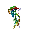
| ||||||||
|---|---|---|---|---|---|---|---|---|---|
| 1 |
| ||||||||
| 単位格子 |
|
-要素
| #1: タンパク質 | / alpha-dystroglycan 分子量: 26226.779 Da / 分子数: 1 / 断片: residues 58-303 / 由来タイプ: 組換発現 / 由来: (組換発現) Mus musculus (ハツカネズミ) / 発現宿主:  Escherichia coli (大腸菌) / 参照: UniProt: Q62165 Escherichia coli (大腸菌) / 参照: UniProt: Q62165 |
|---|---|
| #2: 水 | ChemComp-HOH / 水 分子量: 18.015 Da / 分子数: 199 / 由来タイプ: 天然 / 式: H2O 分子量: 18.015 Da / 分子数: 199 / 由来タイプ: 天然 / 式: H2O |
-実験情報
-実験
| 実験 | 手法: X線回折 / 使用した結晶の数: 1 |
|---|
- 試料調製
試料調製
| 結晶 | マシュー密度: 2.7 Å3/Da / 溶媒含有率: 54.46 % |
|---|
-データ収集
| 放射光源 | 由来: シンクロトロン / サイト: ESRF  / ビームライン: ID14-4 / ビームライン: ID14-4 |
|---|---|
| 検出器 | 検出器: CCD |
| 放射 | プロトコル: SINGLE WAVELENGTH / 単色(M)・ラウエ(L): M / 散乱光タイプ: x-ray |
| 放射波長 | 相対比: 1 |
| 反射 | 解像度: 2.3→35.72 Å / Num. all: 11584 / Num. obs: 11584 / % possible obs: 94.8 % / Observed criterion σ(F): 2 / Observed criterion σ(I): 2 / Biso Wilson estimate: 29 Å2 |
| 反射 シェル | 最高解像度: 2.3 Å / % possible all: 94.8 |
- 解析
解析
| ソフトウェア |
| |||||||||||||||||||||||||
|---|---|---|---|---|---|---|---|---|---|---|---|---|---|---|---|---|---|---|---|---|---|---|---|---|---|---|
| 精密化 | 構造決定の手法: 多重同系置換 / 解像度: 2.3→35.72 Å / Rfactor Rfree error: 0.008 / Data cutoff high absF: 259321.06 / Data cutoff low absF: 0 / Isotropic thermal model: RESTRAINED / 交差検証法: THROUGHOUT / σ(F): 0 / 立体化学のターゲット値: Engh & Huber / 詳細: BULK SOLVENT MODEL USED
| |||||||||||||||||||||||||
| 溶媒の処理 | 溶媒モデル: FLAT MODEL / Bsol: 67.5021 Å2 / ksol: 0.32933 e/Å3 | |||||||||||||||||||||||||
| 原子変位パラメータ | Biso mean: 37.2 Å2
| |||||||||||||||||||||||||
| Refine analyze |
| |||||||||||||||||||||||||
| 精密化ステップ | サイクル: LAST / 解像度: 2.3→35.72 Å
| |||||||||||||||||||||||||
| 拘束条件 |
| |||||||||||||||||||||||||
| LS精密化 シェル | 解像度: 2.3→2.44 Å / Rfactor Rfree error: 0.02 / Total num. of bins used: 6
| |||||||||||||||||||||||||
| Xplor file |
|
