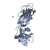
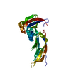
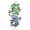
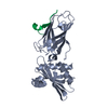
[English] 日本語
Yorodumi
- PDB-2iv8: beta appendage in complex with b-arrestin peptide -

+
Open data


ID or keywords:

Loading...

-
Basic information

Entry
Database: PDB / ID: 2iv8
Titlebeta appendage in complex with b-arrestin peptide
Components
  • AP-2 COMPLEX SUBUNIT BETA-1
  • BETA-ARRESTIN-1Arrestin
KeywordsENDOCYTOSIS/REGULATOR / ENDOCYTOSIS-REGULATOR COMPLEX / 
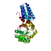
SENSORY TRANSDUCTION / RECEPTOR / COATED PITS / ADAPTOR COMPLEX-REGULATOR
Function / homology
Function and homology information


angiotensin receptor binding / Nef Mediated CD8 Down-regulation / WNT5A-dependent internalization of FZD2, FZD5 and ROR2 / clathrin adaptor complex / Trafficking of GluR2-containing AMPA receptors / WNT5A-dependent internalization of FZD4 / AP-2 adaptor complex / postsynaptic neurotransmitter receptor internalization / Activation of SMO / Retrograde neurotrophin signalling ...angiotensin receptor binding / Nef Mediated CD8 Down-regulation / WNT5A-dependent internalization of FZD2, FZD5 and ROR2 / clathrin adaptor complex / Trafficking of GluR2-containing AMPA receptors / WNT5A-dependent internalization of FZD4 / AP-2 adaptor complex / postsynaptic neurotransmitter receptor internalization / Activation of SMO / Retrograde neurotrophin signalling / negative regulation of interleukin-8 production / clathrin-coated endocytic vesicle / LDL clearance / clathrin-dependent endocytosis / coronary vasculature development / signal sequence binding / arrestin family protein binding / endolysosome membrane / G protein-coupled receptor internalization / Nef Mediated CD4 Down-regulation / enzyme inhibitor activity / aorta development / ventricular septum development / Lysosome Vesicle Biogenesis / positive regulation of Rho protein signal transduction / clathrin binding / Golgi Associated Vesicle Biogenesis / negative regulation of NF-kappaB transcription factor activity / stress fiber assembly / negative regulation of Notch signaling pathway / pseudopodium / Recycling pathway of L1 / negative regulation of interleukin-6 production / positive regulation of receptor internalization / synaptic vesicle endocytosis / EPH-ephrin mediated repulsion of cells / clathrin-coated pit / negative regulation of protein ubiquitination / vesicle-mediated transport / insulin-like growth factor receptor binding / MHC class II antigen presentation / visual perception / VLDLR internalisation and degradation / Activated NOTCH1 Transmits Signal to the Nucleus / GTPase activator activity / kidney development / G protein-coupled receptor binding / intracellular protein transport / clathrin-coated endocytic vesicle membrane / Signaling by high-kinase activity BRAF mutants / MAP2K and MAPK activation / cytoplasmic vesicle membrane / cytoplasmic side of plasma membrane / endocytic vesicle membrane / Signaling by RAF1 mutants / Signaling by moderate kinase activity BRAF mutants / Paradoxical activation of RAF signaling by kinase inactive BRAF / Signaling downstream of RAS mutants / Signaling by BRAF and RAF1 fusions / Thrombin signalling through proteinase activated receptors (PARs) / Cargo recognition for clathrin-mediated endocytosis / protein transport / presynapse / Clathrin-mediated endocytosis / ubiquitin-dependent protein catabolic process / cytoplasmic vesicle / G alpha (s) signalling events / postsynapse / proteasome-mediated ubiquitin-dependent protein catabolic process / Potential therapeutics for SARS / transcription coactivator activity / positive regulation of ERK1 and ERK2 cascade / protein ubiquitination / nuclear body / Ub-specific processing proteases / positive regulation of protein phosphorylation / lysosomal membrane / Golgi membrane / glutamatergic synapse / ubiquitin protein ligase binding / chromatin / regulation of transcription by RNA polymerase II / signal transduction / positive regulation of transcription by RNA polymerase II / nucleoplasm / membrane / nucleus / plasma membrane / cytosol / cytoplasm
Similarity search - Function
Immunoglobulin-like - #1150 / TATA-Binding Protein / Arrestin, conserved site / Arrestins signature. / Arrestin / Arrestin, N-terminal / Clathrin adaptor, beta-adaptin, appendage, Ig-like subdomain / Beta-adaptin appendage, C-terminal subdomain / AP-1/2/4 complex subunit beta / Beta2-adaptin appendage, C-terminal sub-domain ...Immunoglobulin-like - #1150 / TATA-Binding Protein / Arrestin, conserved site / Arrestins signature. / Arrestin / Arrestin, N-terminal / Clathrin adaptor, beta-adaptin, appendage, Ig-like subdomain / Beta-adaptin appendage, C-terminal subdomain / AP-1/2/4 complex subunit beta / Beta2-adaptin appendage, C-terminal sub-domain / Beta2-adaptin appendage, C-terminal sub-domain / AP complex subunit beta / Arrestin-like, N-terminal / Arrestin C-terminal-like domain / Arrestin (or S-antigen), N-terminal domain / Arrestin (or S-antigen), C-terminal domain / Arrestin (or S-antigen), C-terminal domain / Coatomer/calthrin adaptor appendage, C-terminal subdomain / Arrestin-like, C-terminal / Clathrin adaptor, alpha/beta/gamma-adaptin, appendage, Ig-like subdomain / Adaptin C-terminal domain / Adaptin C-terminal domain / Clathrin/coatomer adaptor, adaptin-like, N-terminal / Adaptin N terminal region / Clathrin adaptor, appendage, Ig-like subdomain superfamily / TATA-Binding Protein / TBP domain superfamily / Armadillo/beta-catenin-like repeats / Armadillo / Armadillo-like helical / Immunoglobulin E-set / Armadillo-type fold / Immunoglobulin-like / Sandwich / 2-Layer Sandwich / Mainly Beta / Alpha Beta
Similarity search - Domain/homology
Beta-arrestin-1 / AP-2 complex subunit beta
Similarity search - Component
Biological speciesHOMO SAPIENS (human)
MethodX-RAY DIFFRACTION / SYNCHROTRON / MOLECULAR REPLACEMENT / Resolution: 2.8 Å
AuthorsFord, M.G.J. / Schmid, E.M. / McMahon, H.T.
CitationJournal: Plos Biol. / Year: 2006
Title: Role of the Ap2 Beta-Appendage Hub in Recruiting Partners for Clathrin-Coated Vesicle Assembly.
Authors: Schmid, E.M. / Ford, M.G.J. / Burtey, A. / Praefcke, G.J.K. / Peak-Chew, S. / Mills, I.G. / Benmerah, A. / Mcmahon, H.T.
History
DepositionJun 8, 2006Deposition site: PDBE / Processing site: PDBE
Revision 1.0Jun 12, 2007Provider: repository / Type: Initial release
Revision 1.1Jul 13, 2011Group: Advisory / Version format compliance
Revision 1.2Dec 13, 2023Group: Data collection / Database references ...Data collection / Database references / Other / Refinement description
Category: chem_comp_atom / chem_comp_bond ...chem_comp_atom / chem_comp_bond / database_2 / pdbx_database_status / pdbx_initial_refinement_model
Item: _database_2.pdbx_DOI / _database_2.pdbx_database_accession / _pdbx_database_status.status_code_sf
Remark 700 SHEET THE SHEET STRUCTURE OF THIS MOLECULE IS BIFURCATED. IN ORDER TO REPRESENT THIS FEATURE IN ... SHEET THE SHEET STRUCTURE OF THIS MOLECULE IS BIFURCATED. IN ORDER TO REPRESENT THIS FEATURE IN THE SHEET RECORDS BELOW, TWO SHEETS ARE DEFINED.

-
Structure visualization

Structure viewerMolecule:
MolmilJmol/JSmol

Downloads & links

-
Assembly

Deposited unit
A: AP-2 COMPLEX SUBUNIT BETA-1
P: BETA-ARRESTIN-1
Q: BETA-ARRESTIN-1


Theoretical massNumber of molelcules
Total (without water)31,7343
Polymers31,7343
Non-polymers00
Water28816
1


  • Idetical with deposited unit
  • defined by author&software
TypeNameSymmetry operationNumber
identity operation1_555x,y,z1
Buried area1690 Å2
ΔGint-8 kcal/mol
Surface area12000 Å2
MethodPISA
Unit cell
Length a, b, c (Å)36.952, 35.484, 190.624
Angle α, β, γ (deg.)90.00, 90.00, 90.00
Int Tables number19
Space group name H-MP212121

-
Components

#1: Protein AP-2 COMPLEX SUBUNIT BETA-1 / BETA2-APPENDAGE / ADAPTER-RELATED PROTEIN COMPLEX 2 BETA-1 SUBUNIT / BETA-ADAPTIN / PLASMA MEMBRANE ...BETA2-APPENDAGE / ADAPTER-RELATED PROTEIN COMPLEX 2 BETA-1 SUBUNIT / BETA-ADAPTIN / PLASMA MEMBRANE ADAPTOR HA2/AP2 ADAPTIN BETA SUBUNIT / CLATHRIN ASSEMBLY PROTEIN COMPLEX 2 BETA LARGE CHAIN / AP105B


Mass: 26899.055 Da / Num. of mol.: 1 / Fragment: RESIDUES 700-937
Source method: isolated from a genetically manipulated source
Source: (gene. exp.) HOMO SAPIENS (human) / Organ: BRAIN / Plasmid: PET-15B / Production host: ESCHERICIA COLI (E. coli) / Strain (production host): BL21 / Variant (production host): PLYSS / References: UniProt: P63010
#2: Protein/peptide BETA-ARRESTIN-1 / Arrestin / B-ARRESTIN2


Mass: 2417.651 Da / Num. of mol.: 2 / Fragment: RESIDUES 383-402 / Source method: obtained synthetically / Source: (synth.) HOMO SAPIENS (human) / References: UniProt: P49407
#3: Water ChemComp-HOH / water / Water


Mass: 18.015 Da / Num. of mol.: 16 / Source method: isolated from a natural source / Formula: H2O

-
Experimental details

-
Experiment

ExperimentMethod: X-RAY DIFFRACTION / Number of used crystals: 1

-
Sample preparation

CrystalDensity Matthews: 2.38 Å3/Da / Density % sol: 48 %
Crystal growpH: 7.4
Details: 0.2M MG FORMATE 30% PEG 3350 OTHER REMARKS: THIS WAS THE ONLY CRYSTAL OBTAINED DESPITE HEROIC ATTEMPTS. THE CRYSTAL WAS HIGHLY MOSAIC, AND EPITAXIALLY SPLIT, LIMITING THE AMOUNT OF DATA ...Details: 0.2M MG FORMATE 30% PEG 3350 OTHER REMARKS: THIS WAS THE ONLY CRYSTAL OBTAINED DESPITE HEROIC ATTEMPTS. THE CRYSTAL WAS HIGHLY MOSAIC, AND EPITAXIALLY SPLIT, LIMITING THE AMOUNT OF DATA WHICH COULD BE COLLECTED., pH 7.40

-
Data collection

DiffractionMean temperature: 100 K
Diffraction sourceSource: SYNCHROTRON / Site: ESRF / Beamline: ID14-1 / Wavelength: 0.934
DetectorType: ADSC CCD / Detector: CCD / Date: Sep 1, 2005
RadiationProtocol: SINGLE WAVELENGTH / Monochromatic (M) / Laue (L): M / Scattering type: x-ray
Radiation wavelengthWavelength: 0.934 Å / Relative weight: 1
ReflectionResolution: 2.8→19.78 Å / Num. obs: 4759 / % possible obs: 70.5 % / Redundancy: 5 % / Rmerge(I) obs: 0.06 / Net I/σ(I): 8.7
Reflection shellResolution: 2.8→2.95 Å / Redundancy: 5.4 % / Rmerge(I) obs: 0.15 / Mean I/σ(I) obs: 5 / % possible all: 63.5

-
Processing

Software
NameVersionClassification
REFMAC5.2.0019refinement
MOSFLMdata reduction
SCALAdata scaling
PHASERphasing
RefinementMethod to determine structure: MOLECULAR REPLACEMENT
Starting model: PDB ENTRY 1E42, CHAIN A
Resolution: 2.8→19.78 Å / Cor.coef. Fo:Fc: 0.927 / Cor.coef. Fo:Fc free: 0.781 / SU B: 23.134 / SU ML: 0.455 / TLS residual ADP flag: LIKELY RESIDUAL / Cross valid method: THROUGHOUT / ESU R Free: 0.788 / Stereochemistry target values: MAXIMUM LIKELIHOOD / Details: HYDROGENS HAVE BEEN ADDED IN THE RIDING POSITIONS.
RfactorNum. reflection% reflectionSelection details
Rfree0.369 215 4.5 %RANDOM
Rwork0.213 ---
obs0.22 4512 70.8 %-
Solvent computationIon probe radii: 0.8 Å / Shrinkage radii: 0.8 Å / VDW probe radii: 1.4 Å / Solvent model: MASK
Displacement parametersBiso mean: 33.86 Å2
Baniso -1Baniso -2Baniso -3
1-0.02 Å20 Å20 Å2
2--0.04 Å20 Å2
3----0.06 Å2
Refinement stepCycle: LAST / Resolution: 2.8→19.78 Å
ProteinNucleic acidLigandSolventTotal
Num. atoms2002 0 0 16 2018
Refine LS restraints
Refine-IDTypeDev idealDev ideal targetNumber
X-RAY DIFFRACTIONr_bond_refined_d0.0230.0222058
X-RAY DIFFRACTIONr_bond_other_d
X-RAY DIFFRACTIONr_angle_refined_deg2.2461.9582797
X-RAY DIFFRACTIONr_angle_other_deg
X-RAY DIFFRACTIONr_dihedral_angle_1_deg9.7295251
X-RAY DIFFRACTIONr_dihedral_angle_2_deg44.23725.5198
X-RAY DIFFRACTIONr_dihedral_angle_3_deg23.01515359
X-RAY DIFFRACTIONr_dihedral_angle_4_deg22.835157
X-RAY DIFFRACTIONr_chiral_restr0.1330.2316
X-RAY DIFFRACTIONr_gen_planes_refined0.0070.021558
X-RAY DIFFRACTIONr_gen_planes_other
X-RAY DIFFRACTIONr_nbd_refined0.3050.21033
X-RAY DIFFRACTIONr_nbd_other
X-RAY DIFFRACTIONr_nbtor_refined0.3410.21376
X-RAY DIFFRACTIONr_nbtor_other
X-RAY DIFFRACTIONr_xyhbond_nbd_refined0.2670.296
X-RAY DIFFRACTIONr_xyhbond_nbd_other
X-RAY DIFFRACTIONr_metal_ion_refined
X-RAY DIFFRACTIONr_metal_ion_other
X-RAY DIFFRACTIONr_symmetry_vdw_refined0.3490.237
X-RAY DIFFRACTIONr_symmetry_vdw_other
X-RAY DIFFRACTIONr_symmetry_hbond_refined0.1870.21
X-RAY DIFFRACTIONr_symmetry_hbond_other
X-RAY DIFFRACTIONr_symmetry_metal_ion_refined
X-RAY DIFFRACTIONr_symmetry_metal_ion_other
X-RAY DIFFRACTIONr_mcbond_it0.9731.51301
X-RAY DIFFRACTIONr_mcbond_other
X-RAY DIFFRACTIONr_mcangle_it1.69122048
X-RAY DIFFRACTIONr_mcangle_other
X-RAY DIFFRACTIONr_scbond_it2.2753873
X-RAY DIFFRACTIONr_scbond_other
X-RAY DIFFRACTIONr_scangle_it3.7484.5747
X-RAY DIFFRACTIONr_scangle_other
X-RAY DIFFRACTIONr_long_range_B_refined
X-RAY DIFFRACTIONr_long_range_B_other
X-RAY DIFFRACTIONr_rigid_bond_restr
X-RAY DIFFRACTIONr_sphericity_free
X-RAY DIFFRACTIONr_sphericity_bonded
LS refinement shellResolution: 2.8→2.87 Å / Total num. of bins used: 20 /
RfactorNum. reflection
Rfree0.566 6
Rwork0.238 289
Refinement TLS params.Method: refined / Origin x: -4.9825 Å / Origin y: -4.053 Å / Origin z: -11.088 Å
111213212223313233
T-0.0988 Å2-0.0428 Å2-0.0107 Å2-0.0486 Å2-0.0354 Å2---0.1143 Å2
L0.2184 °2-0.1097 °2-0.7835 °2-0.0579 °20.4901 °2--6.0894 °2
S-0.3225 Å °0.0706 Å °0.0376 Å °0.1401 Å °0.0628 Å °-0.2298 Å °0.1961 Å °-0.3487 Å °0.2597 Å °
Refinement TLS group
IDRefine-IDRefine TLS-IDAuth asym-IDAuth seq-ID
1X-RAY DIFFRACTION1A828 - 905
2X-RAY DIFFRACTION1A906 - 937
3X-RAY DIFFRACTION1P1 - 14

+
About Yorodumi

-
News

-
Feb 9, 2022. New format data for meta-information of EMDB entries

New format data for meta-information of EMDB entries

  • Version 3 of the EMDB header file is now the official format.
  • The previous official version 1.9 will be removed from the archive.

Related info.:EMDB header

External links:wwPDB to switch to version 3 of the EMDB data model

-
Aug 12, 2020. Covid-19 info

Covid-19 info

URL: https://pdbj.org/emnavi/covid19.php

New page: Covid-19 featured information page in EM Navigator.

Related info.:Covid-19 info / Mar 5, 2020. Novel coronavirus structure data

+
Mar 5, 2020. Novel coronavirus structure data

Novel coronavirus structure data

Related info.:Yorodumi Speices / Aug 12, 2020. Covid-19 info

External links:COVID-19 featured content - PDBj / Molecule of the Month (242):Coronavirus Proteases

+
Jan 31, 2019. EMDB accession codes are about to change! (news from PDBe EMDB page)

EMDB accession codes are about to change! (news from PDBe EMDB page)

  • The allocation of 4 digits for EMDB accession codes will soon come to an end. Whilst these codes will remain in use, new EMDB accession codes will include an additional digit and will expand incrementally as the available range of codes is exhausted. The current 4-digit format prefixed with “EMD-” (i.e. EMD-XXXX) will advance to a 5-digit format (i.e. EMD-XXXXX), and so on. It is currently estimated that the 4-digit codes will be depleted around Spring 2019, at which point the 5-digit format will come into force.
  • The EM Navigator/Yorodumi systems omit the EMD- prefix.

Related info.:Q: What is EMD? / ID/Accession-code notation in Yorodumi/EM Navigator

External links:EMDB Accession Codes are Changing Soon! / Contact to PDBj

+
Jul 12, 2017. Major update of PDB

Major update of PDB

  • wwPDB released updated PDB data conforming to the new PDBx/mmCIF dictionary.
  • This is a major update changing the version number from 4 to 5, and with Remediation, in which all the entries are updated.
  • In this update, many items about electron microscopy experimental information are reorganized (e.g. em_software).
  • Now, EM Navigator and Yorodumi are based on the updated data.

External links:wwPDB Remediation / Enriched Model Files Conforming to OneDep Data Standards Now Available in the PDB FTP Archive

-
Yorodumi

Thousand views of thousand structures

  • Yorodumi is a browser for structure data from EMDB, PDB, SASBDB, etc.
  • This page is also the successor to EM Navigator detail page, and also detail information page/front-end page for Omokage search.
  • The word "yorodu" (or yorozu) is an old Japanese word meaning "ten thousand". "mi" (miru) is to see.

Related info.:EMDB / PDB / SASBDB / Comparison of 3 databanks / Yorodumi Search / Aug 31, 2016. New EM Navigator & Yorodumi / Yorodumi Papers / Jmol/JSmol / Function and homology information / Changes in new EM Navigator and Yorodumi

Read more