 Movie
Movie Controller


+ Open data

- Basic information
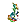
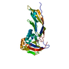
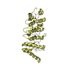
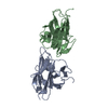
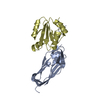
| Entry | Database: PDB / ID: 1u2c | ||||||
|---|---|---|---|---|---|---|---|
| Title | Crystal Structure of a-dystroglycan | ||||||
 Components Components | Dystroglycan | ||||||
 Keywords Keywords | PROTEIN BINDING / IG-like domain / S6 like fold | ||||||
| Function / homology |  Function and homology informationO-linked glycosylation / dystroglycan complex / nerve maturation / muscle attachment / positive regulation of basement membrane assembly involved in embryonic body morphogenesis / retrograde trans-synaptic signaling by trans-synaptic protein complex / contractile ring / Regulation of expression of SLITs and ROBOs / regulation of gastrulation / microtubule anchoring ...O-linked glycosylation / dystroglycan complex / nerve maturation / muscle attachment / positive regulation of basement membrane assembly involved in embryonic body morphogenesis / retrograde trans-synaptic signaling by trans-synaptic protein complex / contractile ring / Regulation of expression of SLITs and ROBOs / regulation of gastrulation / microtubule anchoring / calcium-dependent cell-matrix adhesion / morphogenesis of an epithelial sheet / dystrophin-associated glycoprotein complex / laminin-1 binding / response to denervation involved in regulation of muscle adaptation / basement membrane organization / positive regulation of myelination / regulation of epithelial to mesenchymal transition / skeletal muscle tissue regeneration / photoreceptor ribbon synapse / dystroglycan binding / cellular response to cholesterol / nerve development / vinculin binding / myelination in peripheral nervous system / branching involved in salivary gland morphogenesis / node of Ranvier / costamere / commissural neuron axon guidance / angiogenesis involved in wound healing / response to muscle activity / regulation of neurotransmitter receptor localization to postsynaptic specialization membrane / postsynaptic cytosol / axon regeneration / structural constituent of muscle / positive regulation of cell-matrix adhesion / epithelial tube branching involved in lung morphogenesis / positive regulation of oligodendrocyte differentiation / regulation of synapse organization / negative regulation of MAPK cascade / alpha-actinin binding / plasma membrane raft / membrane protein ectodomain proteolysis / cellular response to organic cyclic compound / basement membrane / negative regulation of phosphatidylinositol 3-kinase/protein kinase B signal transduction / GABA-ergic synapse / laminin binding / Schwann cell development / heart morphogenesis / SH2 domain binding / tubulin binding / nuclear periphery / negative regulation of cell migration / filopodium / axon guidance / morphogenesis of an epithelium / adherens junction / regulation of synaptic plasticity / sarcolemma / response to peptide hormone / cellular response to mechanical stimulus / cell-cell junction / protein transport / virus receptor activity / lamellipodium / actin binding / heart development / postsynapse / postsynaptic membrane / basolateral plasma membrane / cytoskeleton / membrane raft / external side of plasma membrane / focal adhesion / intracellular membrane-bounded organelle / glutamatergic synapse / calcium ion binding / protein-containing complex binding / cell surface / extracellular space / extracellular region / nucleoplasm / membrane / plasma membrane / cytoplasm Function and homology informationO-linked glycosylation / dystroglycan complex / nerve maturation / muscle attachment / positive regulation of basement membrane assembly involved in embryonic body morphogenesis / retrograde trans-synaptic signaling by trans-synaptic protein complex / contractile ring / Regulation of expression of SLITs and ROBOs / regulation of gastrulation / microtubule anchoring ...O-linked glycosylation / dystroglycan complex / nerve maturation / muscle attachment / positive regulation of basement membrane assembly involved in embryonic body morphogenesis / retrograde trans-synaptic signaling by trans-synaptic protein complex / contractile ring / Regulation of expression of SLITs and ROBOs / regulation of gastrulation / microtubule anchoring / calcium-dependent cell-matrix adhesion / morphogenesis of an epithelial sheet / dystrophin-associated glycoprotein complex / laminin-1 binding / response to denervation involved in regulation of muscle adaptation / basement membrane organization / positive regulation of myelination / regulation of epithelial to mesenchymal transition / skeletal muscle tissue regeneration / photoreceptor ribbon synapse / dystroglycan binding / cellular response to cholesterol / nerve development / vinculin binding / myelination in peripheral nervous system / branching involved in salivary gland morphogenesis / node of Ranvier / costamere / commissural neuron axon guidance / angiogenesis involved in wound healing / response to muscle activity / regulation of neurotransmitter receptor localization to postsynaptic specialization membrane / postsynaptic cytosol / axon regeneration / structural constituent of muscle / positive regulation of cell-matrix adhesion / epithelial tube branching involved in lung morphogenesis / positive regulation of oligodendrocyte differentiation / regulation of synapse organization / negative regulation of MAPK cascade / alpha-actinin binding / plasma membrane raft / membrane protein ectodomain proteolysis / cellular response to organic cyclic compound / basement membrane / negative regulation of phosphatidylinositol 3-kinase/protein kinase B signal transduction / GABA-ergic synapse / laminin binding / Schwann cell development / heart morphogenesis / SH2 domain binding / tubulin binding / nuclear periphery / negative regulation of cell migration / filopodium / axon guidance / morphogenesis of an epithelium / adherens junction / regulation of synaptic plasticity / sarcolemma / response to peptide hormone / cellular response to mechanical stimulus / cell-cell junction / protein transport / virus receptor activity / lamellipodium / actin binding / heart development / postsynapse / postsynaptic membrane / basolateral plasma membrane / cytoskeleton / membrane raft / external side of plasma membrane / focal adhesion / intracellular membrane-bounded organelle / glutamatergic synapse / calcium ion binding / protein-containing complex binding / cell surface / extracellular space / extracellular region / nucleoplasm / membrane / plasma membrane / cytoplasmSimilarity search - Function | ||||||
| Biological species |  Mus musculus (house mouse) Mus musculus (house mouse) | ||||||
| Method | X-RAY DIFFRACTION / SYNCHROTRON / MIR / Resolution: 2.3 Å | ||||||
 Authors Authors | Bozic, D. / Sciandra, F. / Lamba, D. / Brancaccio, A. | ||||||
 Citation Citation | Journal: J.Biol.Chem. / Year: 2004 Title: The Structure of the N-terminal Region of Murine Skeletal Muscle {alpha}-Dystroglycan Discloses a Modular Architecture Authors: Bozic, D. / Sciandra, F. / Lamba, D. / Brancaccio, A. | ||||||
| History |
|
- Structure visualization
| Structure viewer | Molecule: MolmilJmol/JSmol |
|---|
- Downloads & links
-Download
| PDBx/mmCIF format | 1u2c.cif.gz | 56 KB | Display | PDBx/mmCIF format |
|---|---|---|---|---|
| PDB format | pdb1u2c.ent.gz | 43.6 KB | Display | PDB format |
| PDBx/mmJSON format | 1u2c.json.gz | Tree view | PDBx/mmJSON format | |
| Others |  Other downloads Other downloads |
-Validation report
| Arichive directory | https://data.pdbj.org/pub/pdb/validation_reports/u2/1u2cftp://data.pdbj.org/pub/pdb/validation_reports/u2/1u2c | HTTPS FTP |
|---|
-Related structure data
| Similar structure data |
|---|


-Links
 PDBj
PDBj
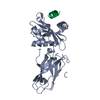
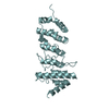
- Assembly
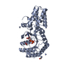
| Deposited unit | 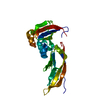
| ||||||||
|---|---|---|---|---|---|---|---|---|---|
| 1 |
| ||||||||
| Unit cell |
|
-Components
| #1: Protein | / alpha-dystroglycan Mass: 26226.779 Da / Num. of mol.: 1 / Fragment: residues 58-303 Source method: isolated from a genetically manipulated source Source: (gene. exp.) Mus musculus (house mouse) / Production host:  Escherichia coli (E. coli) / References: UniProt: Q62165 Escherichia coli (E. coli) / References: UniProt: Q62165 |
|---|---|
| #2: Water | ChemComp-HOH / Water Mass: 18.015 Da / Num. of mol.: 199 / Source method: isolated from a natural source / Formula: H2O Mass: 18.015 Da / Num. of mol.: 199 / Source method: isolated from a natural source / Formula: H2O |
-Experimental details
-Experiment
| Experiment | Method: X-RAY DIFFRACTION / Number of used crystals: 1 |
|---|
- Sample preparation
| Crystal | Density Matthews: 2.7 Å3/Da / Density % sol: 54.46 % |
|---|
-Data collection
| Diffraction source | Source: SYNCHROTRON / Site: ESRF  / Beamline: ID14-4 / Beamline: ID14-4 |
|---|---|
| Detector | Detector: CCD |
| Radiation | Protocol: SINGLE WAVELENGTH / Monochromatic (M) / Laue (L): M / Scattering type: x-ray |
| Radiation wavelength | Relative weight: 1 |
| Reflection | Resolution: 2.3→35.72 Å / Num. all: 11584 / Num. obs: 11584 / % possible obs: 94.8 % / Observed criterion σ(F): 2 / Observed criterion σ(I): 2 / Biso Wilson estimate: 29 Å2 |
| Reflection shell | Highest resolution: 2.3 Å / % possible all: 94.8 |
- Processing
| Software |
| |||||||||||||||||||||||||
|---|---|---|---|---|---|---|---|---|---|---|---|---|---|---|---|---|---|---|---|---|---|---|---|---|---|---|
| Refinement | Method to determine structure: MIR / Resolution: 2.3→35.72 Å / Rfactor Rfree error: 0.008 / Data cutoff high absF: 259321.06 / Data cutoff low absF: 0 / Isotropic thermal model: RESTRAINED / Cross valid method: THROUGHOUT / σ(F): 0 / Stereochemistry target values: Engh & Huber / Details: BULK SOLVENT MODEL USED
| |||||||||||||||||||||||||
| Solvent computation | Solvent model: FLAT MODEL / Bsol: 67.5021 Å2 / ksol: 0.32933 e/Å3 | |||||||||||||||||||||||||
| Displacement parameters | Biso mean: 37.2 Å2
| |||||||||||||||||||||||||
| Refine analyze |
| |||||||||||||||||||||||||
| Refinement step | Cycle: LAST / Resolution: 2.3→35.72 Å
| |||||||||||||||||||||||||
| Refine LS restraints |
| |||||||||||||||||||||||||
| LS refinement shell | Resolution: 2.3→2.44 Å / Rfactor Rfree error: 0.02 / Total num. of bins used: 6
| |||||||||||||||||||||||||
| Xplor file |
|
