 Movie
Movie Controller

[English] 日本語
 Yorodumi
- PDB-1tuk: Crystal structure of liganded type 2 non specific lipid transfer ... -
+ Open data

- Basic information
| Entry | Database: PDB / ID: 1tuk | ||||||
|---|---|---|---|---|---|---|---|
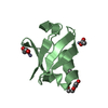
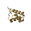
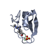
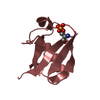
| Title | Crystal structure of liganded type 2 non specific lipid transfer protein from wheat | ||||||
 Components Components | Nonspecific lipid-transfer protein 2G | ||||||
 Keywords Keywords | LIPID TRANSPORT / ns-LTP2 /  lipid transfer protein lipid transfer protein | ||||||
| Function / homology |  Function and homology information Function and homology informationlipid transport / cell wall organization / lipid binding / extracellular regionSimilarity search - Function | ||||||
| Biological species |  Triticum aestivum (bread wheat) Triticum aestivum (bread wheat) | ||||||
| Method | X-RAY DIFFRACTION / SYNCHROTRON / AB INITIO PHASING / Resolution: 1.12 Å | ||||||
 Authors Authors | Hoh, F. / Pons, J.L. / Gautier, M.F. / De Lamotte, F. / Dumas, C. | ||||||
 Citation Citation | Journal: Acta Crystallogr.,Sect.D / Year: 2005 Title: Structure of a liganded type 2 non-specific lipid-transfer protein from wheat and the molecular basis of lipid binding. Authors: Hoh, F. / Pons, J.L. / Gautier, M.F. / de Lamotte, F. / Dumas, C. #1: Journal: J.BIOL.CHEM. / Year: 2003Title: Refined solution structure of a liganded type 2 wheat nonspecific lipid transfer protein Authors: Pons, J.L. / de Lamotte, F. / Gautier, M.F. / Delsuc, M.A. | ||||||
| History |
|
- Structure visualization
| Structure viewer | Molecule: MolmilJmol/JSmol |
|---|
- Downloads & links
-Download
| PDBx/mmCIF format | 1tuk.cif.gz | 37.3 KB | Display | PDBx/mmCIF format |
|---|---|---|---|---|
| PDB format | pdb1tuk.ent.gz | 29.2 KB | Display | PDB format |
| PDBx/mmJSON format | 1tuk.json.gz | Tree view | PDBx/mmJSON format | |
| Others |  Other downloads Other downloads |
-Validation report
| Arichive directory | https://data.pdbj.org/pub/pdb/validation_reports/tu/1tukftp://data.pdbj.org/pub/pdb/validation_reports/tu/1tuk | HTTPS FTP |
|---|
-Related structure data
| Related structure data | |
|---|---|
| Similar structure data |


-Links
 PDBj
PDBj
- Assembly
| Deposited unit | 
| ||||||||
|---|---|---|---|---|---|---|---|---|---|
| 1 |
| ||||||||
| Unit cell |
|
-Components
| #1: Protein | Mass: 6985.943 Da / Num. of mol.: 1 / Source method: isolated from a natural source / Source: (natural) Triticum aestivum (bread wheat) / References: UniProt: P82900 | ||||
|---|---|---|---|---|---|
| #2: Chemical | Iodide  Mass: 126.904 Da / Num. of mol.: 2 / Source method: obtained synthetically / Formula: I Mass: 126.904 Da / Num. of mol.: 2 / Source method: obtained synthetically / Formula: I#3: Chemical |   Mass: 483.553 Da / Num. of mol.: 2 / Source method: obtained synthetically / Formula: C22H44O9P Mass: 483.553 Da / Num. of mol.: 2 / Source method: obtained synthetically / Formula: C22H44O9P#4: Water | ChemComp-HOH / | Water Mass: 18.015 Da / Num. of mol.: 75 / Source method: isolated from a natural source / Formula: H2O Mass: 18.015 Da / Num. of mol.: 75 / Source method: isolated from a natural source / Formula: H2O |
-Experimental details
-Experiment
| Experiment | Method: X-RAY DIFFRACTION / Number of used crystals: 1 |
|---|
- Sample preparation
| Crystal | Density Matthews: 1.91 Å3/Da / Density % sol: 35.5 % |
|---|---|
| Crystal grow | Temperature: 298 K / Method: vapor diffusion, hanging drop / pH: 6 Details: PEG 6000, VAPOR DIFFUSION, HANGING DROP, temperature 298K |
-Data collection
| Diffraction | Mean temperature: 100 K |
|---|---|
| Diffraction source | Source: SYNCHROTRON / Site: ESRF  / Beamline: BM14 / Wavelength: 0.8 Å / Beamline: BM14 / Wavelength: 0.8 Å |
| Detector | Type: MARRESEARCH / Detector: CCD / Date: Sep 16, 2002 |
| Radiation | Monochromator: Mirror / Protocol: SINGLE WAVELENGTH / Monochromatic (M) / Laue (L): M / Scattering type: x-ray |
| Radiation wavelength | Wavelength: 0.8 Å / Relative weight: 1 |
| Reflection | Resolution: 1.12→32.97 Å / Num. all: 22535 / Num. obs: 22535 / % possible obs: 94.8 % / Observed criterion σ(F): 0 / Observed criterion σ(I): 0 |
| Reflection shell | Resolution: 1.123→1.153 Å / % possible all: 79.71 |
- Processing
| Software |
| ||||||||||||||||||||||||||||||||||||||||||||||||||||||||||||||||||||||||||||||||||||||||||||||||||||||||||||||||||||||||||||||||||||||||||||||||||||||||||||||||||||||||||
|---|---|---|---|---|---|---|---|---|---|---|---|---|---|---|---|---|---|---|---|---|---|---|---|---|---|---|---|---|---|---|---|---|---|---|---|---|---|---|---|---|---|---|---|---|---|---|---|---|---|---|---|---|---|---|---|---|---|---|---|---|---|---|---|---|---|---|---|---|---|---|---|---|---|---|---|---|---|---|---|---|---|---|---|---|---|---|---|---|---|---|---|---|---|---|---|---|---|---|---|---|---|---|---|---|---|---|---|---|---|---|---|---|---|---|---|---|---|---|---|---|---|---|---|---|---|---|---|---|---|---|---|---|---|---|---|---|---|---|---|---|---|---|---|---|---|---|---|---|---|---|---|---|---|---|---|---|---|---|---|---|---|---|---|---|---|---|---|---|---|---|---|
| Refinement | Method to determine structure: AB INITIO PHASING / Resolution: 1.12→32.9 Å / Cor.coef. Fo:Fc: 0.969 / Cor.coef. Fo:Fc free: 0.97 / SU B: 0.855 / SU ML: 0.018 / Cross valid method: THROUGHOUT / σ(F): 0 / ESU R: 0.032 / ESU R Free: 0.029 / Stereochemistry target values: MAXIMUM LIKELIHOOD
| ||||||||||||||||||||||||||||||||||||||||||||||||||||||||||||||||||||||||||||||||||||||||||||||||||||||||||||||||||||||||||||||||||||||||||||||||||||||||||||||||||||||||||
| Solvent computation | Ion probe radii: 0.8 Å / Shrinkage radii: 0.8 Å / VDW probe radii: 1.2 Å / Solvent model: BABINET MODEL WITH MASK | ||||||||||||||||||||||||||||||||||||||||||||||||||||||||||||||||||||||||||||||||||||||||||||||||||||||||||||||||||||||||||||||||||||||||||||||||||||||||||||||||||||||||||
| Displacement parameters | Biso mean: 12.01 Å2
| ||||||||||||||||||||||||||||||||||||||||||||||||||||||||||||||||||||||||||||||||||||||||||||||||||||||||||||||||||||||||||||||||||||||||||||||||||||||||||||||||||||||||||
| Refinement step | Cycle: LAST / Resolution: 1.12→32.9 Å
| ||||||||||||||||||||||||||||||||||||||||||||||||||||||||||||||||||||||||||||||||||||||||||||||||||||||||||||||||||||||||||||||||||||||||||||||||||||||||||||||||||||||||||
| Refine LS restraints |
| ||||||||||||||||||||||||||||||||||||||||||||||||||||||||||||||||||||||||||||||||||||||||||||||||||||||||||||||||||||||||||||||||||||||||||||||||||||||||||||||||||||||||||
| LS refinement shell | Resolution: 1.123→1.153 Å / Total num. of bins used: 20
|
