 Movie
Movie Controller

[English] 日本語
 Yorodumi
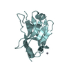
- PDB-1sau: The Gamma subunit of the dissimilatory sulfite reductase (DsrC) f... -
+ Open data

- Basic information
| Entry | Database: PDB / ID: 1sau | ||||||
|---|---|---|---|---|---|---|---|
| Title | The Gamma subunit of the dissimilatory sulfite reductase (DsrC) from Archaeoglobus fulgidus at 1.1 A resolution | ||||||
 Components Components | sulfite reductase, desulfoviridin-type subunit gamma | ||||||
 Keywords Keywords | OXIDOREDUCTASE / ORTHOGONAL HELICAL BUNDLE | ||||||
| Function / homology |  Function and homology information Function and homology informationsulfur carrier activity / tRNA wobble position uridine thiolation / cytoplasmSimilarity search - Function | ||||||
| Biological species |   Archaeoglobus fulgidus (archaea) Archaeoglobus fulgidus (archaea) | ||||||
| Method | X-RAY DIFFRACTION / SYNCHROTRON / SAD / Resolution: 1.12 Å | ||||||
 Authors Authors | Mander, G.J. / Weiss, M.S. / Hedderich, R. / Kahnt, J. / Ermler, U. / Warkentin, E. | ||||||
 Citation Citation | Journal: Acta Crystallogr.,Sect.D / Year: 2004 Title: Determination of a novel structure by a combination of long-wavelength sulfur phasing and radiation-damage-induced phasing. Authors: Weiss, M.S. / Mander, G. / Hedderich, R. / Diederichs, K. / Ermler, U. / Warkentin, E. #1: Journal: Acta Crystallogr.,Sect.D / Year: 2004Title: Determination of a novel structure by a combination of long-wavelength sulfur phasing and radiation-damage-induced phasing. Authors: Weiss, M.S. / Mander, G. / Hedderich, R. / Diederichs, K. / Ermler, U. / Warkentin, E. | ||||||
| History |
|
- Structure visualization
| Structure viewer | Molecule: MolmilJmol/JSmol |
|---|
- Downloads & links
-Download
| PDBx/mmCIF format | 1sau.cif.gz | 65.7 KB | Display | PDBx/mmCIF format |
|---|---|---|---|---|
| PDB format | pdb1sau.ent.gz | 52.9 KB | Display | PDB format |
| PDBx/mmJSON format | 1sau.json.gz | Tree view | PDBx/mmJSON format | |
| Others |  Other downloads Other downloads |
-Validation report
| Arichive directory | https://data.pdbj.org/pub/pdb/validation_reports/sa/1sauftp://data.pdbj.org/pub/pdb/validation_reports/sa/1sau | HTTPS FTP |
|---|
-Related structure data
| Similar structure data |
|---|


-Links
 PDBj
PDBj- Assembly
| Deposited unit | 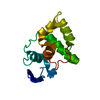
| ||||||||
|---|---|---|---|---|---|---|---|---|---|
| 1 |
| ||||||||
| Unit cell |
|
-Components
| #1: Protein | / DsrC / dissimilatory sulfite reductase Mass: 13498.659 Da / Num. of mol.: 1 Source method: isolated from a genetically manipulated source Source: (gene. exp.) Archaeoglobus fulgidus (archaea) / Strain: DSM 4304 / Gene: DsrC / Plasmid: pCR2.1-TOPO / Production host:  Escherichia coli (E. coli) / Strain (production host): BL21(DE3) / K12 / References: UniProt: O28055 Escherichia coli (E. coli) / Strain (production host): BL21(DE3) / K12 / References: UniProt: O28055 |
|---|---|
| #2: Water | ChemComp-HOH / Water Mass: 18.015 Da / Num. of mol.: 149 / Source method: isolated from a natural source / Formula: H2O Mass: 18.015 Da / Num. of mol.: 149 / Source method: isolated from a natural source / Formula: H2O |
-Experimental details
-Experiment
| Experiment | Method: X-RAY DIFFRACTION / Number of used crystals: 1 |
|---|
- Sample preparation
| Crystal | Density Matthews: 1.8 Å3/Da / Density % sol: 31.23 % | ||||||||||||||||||
|---|---|---|---|---|---|---|---|---|---|---|---|---|---|---|---|---|---|---|---|
| Crystal grow | Temperature: 277 K / pH: 9.5 Details: TRIS, MPD, pH 9.5, VAPOR DIFFUSION, HANGING DROP, temperature 277K, pH 9.50 | ||||||||||||||||||
| Crystal grow | *PLUS pH: 9.5 / Method: vapor diffusion, hanging drop | ||||||||||||||||||
| Components of the solutions | *PLUS
|
-Data collection
| Diffraction | Mean temperature: 100 K |
|---|---|
| Diffraction source | Source: SYNCHROTRON / Site: ESRF  / Beamline: ID14-4 / Wavelength: 0.9393 / Beamline: ID14-4 / Wavelength: 0.9393 |
| Detector | Type: ADSC QUANTUM 4 / Detector: CCD / Date: Jul 11, 2002 / Details: MIRROR |
| Radiation | Monochromator: SI(111) / Protocol: SINGLE WAVELENGTH / Monochromatic (M) / Laue (L): M / Scattering type: x-ray |
| Radiation wavelength | Wavelength: 0.9393 Å / Relative weight: 1 |
| Reflection | Resolution: 1.1→20 Å / Num. obs: 40859 / % possible obs: 93.1 % / Observed criterion σ(I): 0 / Redundancy: 5 % / Biso Wilson estimate: 9.4 Å2 / Rsym value: 0.083 / Net I/σ(I): 21 |
| Reflection shell | Resolution: 1.1→1.18 Å / Redundancy: 2.3 % / Mean I/σ(I) obs: 5.5 / Rsym value: 0.165 / % possible all: 73 |
| Reflection | *PLUS Highest resolution: 1.12 Å / Lowest resolution: 99 Å / Num. obs: 40862 / Observed criterion σ(I): 0 / Redundancy: 5 % / Num. measured all: 205663 / Rmerge(I) obs: 0.093 |
| Reflection shell | *PLUS Highest resolution: 1.12 Å / Lowest resolution: 1.15 Å / % possible obs: 73.5 % / Redundancy: 2.3 % / Rmerge(I) obs: 0.16 / Mean I/σ(I) obs: 4.7 |
- Processing
| Software |
| |||||||||||||||||||||||||||||||||
|---|---|---|---|---|---|---|---|---|---|---|---|---|---|---|---|---|---|---|---|---|---|---|---|---|---|---|---|---|---|---|---|---|---|---|
| Refinement | Method to determine structure: SAD / Resolution: 1.12→20 Å / Num. parameters: 10819 / Num. restraintsaints: 14685 / Cross valid method: THROUGHOUT / σ(F): 0 / Stereochemistry target values: ENGH & HUBER Details: ANISOTROPIC REFINEMENT REDUCED FREE R (NO CUTOFF) BY 0.044
| |||||||||||||||||||||||||||||||||
| Solvent computation | Solvent model: MOEWS & KRETSINGER, J.MOL.BIOL.91(1973)201-228. riding hydrogens were used in refinement. | |||||||||||||||||||||||||||||||||
| Refine analyze | Num. disordered residues: 18 / Occupancy sum hydrogen: 938 / Occupancy sum non hydrogen: 1062.5 | |||||||||||||||||||||||||||||||||
| Refinement step | Cycle: LAST / Resolution: 1.12→20 Å
| |||||||||||||||||||||||||||||||||
| Refine LS restraints |
| |||||||||||||||||||||||||||||||||
| LS refinement shell | Resolution: 1.12→1.17 Å /
| |||||||||||||||||||||||||||||||||
| Software | *PLUS Name: SHELXL / Version: 97 / Classification: refinement | |||||||||||||||||||||||||||||||||
| Refinement | *PLUS Lowest resolution: 30 Å / Rfactor obs: 0.108 / Rfactor Rwork: 0.108 | |||||||||||||||||||||||||||||||||
| Solvent computation | *PLUS | |||||||||||||||||||||||||||||||||
| Displacement parameters | *PLUS Biso mean: 8.2 Å2 | |||||||||||||||||||||||||||||||||
| LS refinement shell | *PLUS Lowest resolution: 1.15 Å |
