 Movie
Movie Controller

[English] 日本語
 Yorodumi
- PDB-1rm4: Crystal structure of recombinant photosynthetic glyceraldehyde-3-... -
+ Open data

- Basic information
| Entry | Database: PDB / ID: 1rm4 | ||||||
|---|---|---|---|---|---|---|---|
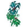
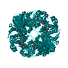
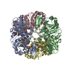
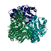
| Title | Crystal structure of recombinant photosynthetic glyceraldehyde-3-phosphate dehydrogenase A4 isoform, complexed with NADP | ||||||
 Components Components | Glyceraldehyde 3-phosphate dehydrogenase A | ||||||
 Keywords Keywords |  OXIDOREDUCTASE / Rossmann fold / GAPDH-NADP complex OXIDOREDUCTASE / Rossmann fold / GAPDH-NADP complex | ||||||
| Function / homology |  Function and homology informationglyceraldehyde-3-phosphate dehydrogenase (NADP+) (phosphorylating) / glyceraldehyde-3-phosphate dehydrogenase (NADP+) (phosphorylating) activity / reductive pentose-phosphate cycle / chloroplast / glucose metabolic process / NAD binding / NADP binding Function and homology informationglyceraldehyde-3-phosphate dehydrogenase (NADP+) (phosphorylating) / glyceraldehyde-3-phosphate dehydrogenase (NADP+) (phosphorylating) activity / reductive pentose-phosphate cycle / chloroplast / glucose metabolic process / NAD binding / NADP bindingSimilarity search - Function | ||||||
| Biological species |  Spinacia oleracea (spinach) Spinacia oleracea (spinach) | ||||||
| Method | X-RAY DIFFRACTION / SYNCHROTRON / MOLECULAR REPLACEMENT / Resolution: 2 Å | ||||||
 Authors Authors | Sparla, F. / Fermani, S. / Falini, G. / Ripamonti, A. / Sabatino, P. / Pupillo, P. / Trost, P. | ||||||
 Citation Citation | Journal: J.Mol.Biol. / Year: 2004 Title: Coenzyme Site-directed Mutants of Photosynthetic A(4)-GAPDH Show Selectively Reduced NADPH-dependent Catalysis, Similar to Regulatory AB-GAPDH Inhibited by Oxidized Thioredoxin Authors: Sparla, F. / Fermani, S. / Falini, G. / Zaffagnini, M. / Ripamonti, A. / Sabatino, P. / Pupillo, P. / Trost, P. #1: Journal: Biochemistry / Year: 2003Title: The dual coenzyme specificity of photosynthetic glyceraldehyde-3-phosphate dehydrogenase interpreted by the crystal structure of A4 isoform complexed with NAD Authors: Falini, G. / Fermani, S. / Ripamonti, A. / Sabatino, P. / Sparla, F. / Pupillo, P. / Trost, P. #2: Journal: J.Mol.Biol. / Year: 2001Title: Crystal structure of the non-regulatory A4 isoform of spinach chloroplast glyceraldehyde-3-phosphate dehydrogenase complexed with NADP Authors: Fermani, S. / Ripamonti, A. / Sabatino, P. / Zanotti, G. / Scagliarini, S. / Sparla, F. / Trost, P. / Pupillo, P. | ||||||
| History |
|
- Structure visualization
| Structure viewer | Molecule: MolmilJmol/JSmol |
|---|
- Downloads & links
-Download
| PDBx/mmCIF format | 1rm4.cif.gz | 219 KB | Display | PDBx/mmCIF format |
|---|---|---|---|---|
| PDB format | pdb1rm4.ent.gz | 174.9 KB | Display | PDB format |
| PDBx/mmJSON format | 1rm4.json.gz | Tree view | PDBx/mmJSON format | |
| Others |  Other downloads Other downloads |
-Validation report
| Arichive directory | https://data.pdbj.org/pub/pdb/validation_reports/rm/1rm4ftp://data.pdbj.org/pub/pdb/validation_reports/rm/1rm4 | HTTPS FTP |
|---|
-Related structure data
| Related structure data |  1rm3C 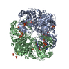 1rm5C  1jn0S S: Starting model for refinement C: citing same article ( |
|---|---|
| Similar structure data |


-Links
 PDBj
PDBj


- Assembly
| Deposited unit | 
| |||||||||||||||
|---|---|---|---|---|---|---|---|---|---|---|---|---|---|---|---|---|
| 1 | 
| |||||||||||||||
| 2 | 
| |||||||||||||||
| Unit cell |
| |||||||||||||||
| Components on special symmetry positions |
| |||||||||||||||
| Details | The biological assembly is a tetramer (OPQR) generated from the monomer O by the operations: monomer R -x, y, -z and translation 0, 0, 2; monomer Q x, -y, -z and translation 0, 1, 2; monomer P -x, -y, z and translation 0, 1, 0. A second tetramer is generated from dimer AB by the operations: 1/2-x, 3/2-y, z and translation 0, 0, 0. |
-Components
| #1: Protein | Mass: 36256.391 Da / Num. of mol.: 3 Source method: isolated from a genetically manipulated source Source: (gene. exp.) Spinacia oleracea (spinach) / Gene: GapA / Organ: leaves / Organelle: chloroplastsChloroplast / Plasmid: PET-28 / Species (production host): Escherichia coli / Production host:  Escherichia coli BL21(DE3) (bacteria) / Strain (production host): BL21(DE3) Escherichia coli BL21(DE3) (bacteria) / Strain (production host): BL21(DE3)References: UniProt: P19866, glyceraldehyde-3-phosphate dehydrogenase (NADP+) (phosphorylating)#2: Chemical | ChemComp-SO4 / Sulfate  Mass: 96.063 Da / Num. of mol.: 8 / Source method: obtained synthetically / Formula: SO4 Mass: 96.063 Da / Num. of mol.: 8 / Source method: obtained synthetically / Formula: SO4#3: Chemical | Nicotinamide adenine dinucleotide phosphate  Mass: 745.421 Da / Num. of mol.: 3 / Source method: obtained synthetically / Formula: C21H30N7O17P3 Mass: 745.421 Da / Num. of mol.: 3 / Source method: obtained synthetically / Formula: C21H30N7O17P3#4: Water | ChemComp-HOH / | Water Mass: 18.015 Da / Num. of mol.: 589 / Source method: isolated from a natural source / Formula: H2O Mass: 18.015 Da / Num. of mol.: 589 / Source method: isolated from a natural source / Formula: H2O |
|---|
-Experimental details
-Experiment
| Experiment | Method: X-RAY DIFFRACTION / Number of used crystals: 1 |
|---|
- Sample preparation
| Crystal | Density Matthews: 2.96 Å3/Da / Density % sol: 58.1 % |
|---|---|
| Crystal grow | Temperature: 293 K / Method: vapor diffusion, hanging drop / pH: 8.5 Details: ammonium sulphate, Tris-HCl, pH 8.5, VAPOR DIFFUSION, HANGING DROP, temperature 293K |
-Data collection
| Diffraction | Mean temperature: 100 K |
|---|---|
| Diffraction source | Source: SYNCHROTRON / Site: ELETTRA  / Beamline: 5.2R / Wavelength: 1 Å / Beamline: 5.2R / Wavelength: 1 Å |
| Detector | Type: MARRESEARCH / Detector: CCD / Date: Mar 19, 2001 / Details: mirrors |
| Radiation | Monochromator: Si crystal / Protocol: SINGLE WAVELENGTH / Monochromatic (M) / Laue (L): M / Scattering type: x-ray |
| Radiation wavelength | Wavelength: 1 Å / Relative weight: 1 |
| Reflection | Resolution: 2→76 Å / Num. all: 94456 / Num. obs: 81938 / % possible obs: 52 % / Observed criterion σ(I): -3 / Redundancy: 5 % / Biso Wilson estimate: 14.2 Å2 / Rsym value: 0.038 / Net I/σ(I): 24.2 |
| Reflection shell | Resolution: 2→2.07 Å / Mean I/σ(I) obs: 3.8 / Num. unique all: 7906 / Rsym value: 0.115 / % possible all: 37.6 |
- Processing
| Software |
| ||||||||||||||||||||||||||||||||||||
|---|---|---|---|---|---|---|---|---|---|---|---|---|---|---|---|---|---|---|---|---|---|---|---|---|---|---|---|---|---|---|---|---|---|---|---|---|---|
| Refinement | Method to determine structure: MOLECULAR REPLACEMENT Starting model: 1JN0 Resolution: 2→69.95 Å / Rfactor Rfree error: 0.004 / Data cutoff high absF: 4753767.43 / Data cutoff low absF: 0 / Isotropic thermal model: RESTRAINED / Cross valid method: THROUGHOUT / σ(F): 0 / Stereochemistry target values: Engh & Huber
| ||||||||||||||||||||||||||||||||||||
| Solvent computation | Solvent model: FLAT MODEL / Bsol: 63.3863 Å2 / ksol: 0.394412 e/Å3 | ||||||||||||||||||||||||||||||||||||
| Displacement parameters | Biso mean: 32 Å2
| ||||||||||||||||||||||||||||||||||||
| Refine analyze |
| ||||||||||||||||||||||||||||||||||||
| Refinement step | Cycle: LAST / Resolution: 2→69.95 Å
| ||||||||||||||||||||||||||||||||||||
| Refine LS restraints |
| ||||||||||||||||||||||||||||||||||||
| LS refinement shell | Resolution: 2→2.13 Å / Rfactor Rfree error: 0.011 / Total num. of bins used: 6
| ||||||||||||||||||||||||||||||||||||
| Xplor file |
|
