 Movie
Movie Controller

[English] 日本語
 Yorodumi
- PDB-1jn0: Crystal structure of the non-regulatory A4 isoform of spinach chl... -
+ Open data

- Basic information
| Entry | Database: PDB / ID: 1jn0 | ||||||
|---|---|---|---|---|---|---|---|
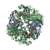
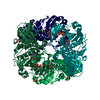
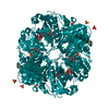
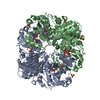
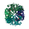
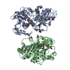
| Title | Crystal structure of the non-regulatory A4 isoform of spinach chloroplast glyceraldehyde-3-phosphate dehydrogenase complexed with NADP | ||||||
 Components Components | GLYCERALDEHYDE-3-PHOSPHATE DEHYDROGENASE A | ||||||
 Keywords Keywords |  OXIDOREDUCTASE / ROSSMANN FOLD / PROTEIN-NADP COMPLEX / NADPH OXIDOREDUCTASE / ROSSMANN FOLD / PROTEIN-NADP COMPLEX / NADPH | ||||||
| Function / homology |  Function and homology informationglyceraldehyde-3-phosphate dehydrogenase (NADP+) (phosphorylating) / glyceraldehyde-3-phosphate dehydrogenase (NADP+) (phosphorylating) activity / reductive pentose-phosphate cycle / chloroplast / glucose metabolic process / NAD binding / NADP binding Function and homology informationglyceraldehyde-3-phosphate dehydrogenase (NADP+) (phosphorylating) / glyceraldehyde-3-phosphate dehydrogenase (NADP+) (phosphorylating) activity / reductive pentose-phosphate cycle / chloroplast / glucose metabolic process / NAD binding / NADP bindingSimilarity search - Function | ||||||
| Biological species |  Spinacia oleracea (spinach) Spinacia oleracea (spinach) | ||||||
| Method | X-RAY DIFFRACTION / SYNCHROTRON / MOLECULAR REPLACEMENT / Resolution: 3 Å | ||||||
 Authors Authors | Fermani, S. / Ripamonti, A. / Sabatino, P. / Zanotti, G. / Scagliarini, S. / Sparla, F. / Trost, P. / Pupillo, P. | ||||||
 Citation Citation | Journal: J.Mol.Biol. / Year: 2001 Title: Crystal structure of the non-regulatory A(4 )isoform of spinach chloroplast glyceraldehyde-3-phosphate dehydrogenase complexed with NADP. Authors: Fermani, S. / Ripamonti, A. / Sabatino, P. / Zanotti, G. / Scagliarini, S. / Sparla, F. / Trost, P. / Pupillo, P. #1: Journal: Acta Crystallogr.,Sect.D / Year: 1999Title: Crystallization and preliminary X-ray study of chloroplast glyceraldehyde-3-phosphate dehydrogenase Authors: Sabatino, P. / Fermani, S. / Ripamonti, A. / Cassetta, A. / Scagliarini, S. / Trost, P. #2: Journal: J.Exp.Bot. / Year: 1998Title: The non-regulatory isoform of NADP(H)-glyceraldehyde-3-phosphate dehydrogenase from spinach chloroplasts Authors: Scagliarini, S. / Trost, P. / Pupillo, P. #3: Journal: Biochim.Biophys.Acta / Year: 1990Title: Chloroplast glyceraldehyde-3-phosphate dehydrogenase (NADP): amino acid sequence of the subunits from isoenzyme I and structural relationship with isoenzyme II. Authors: Ferri, G. / Stoppini, M. / Meloni, M. / Zapponi, M.C. / Iadarola, P. | ||||||
| History |
|
- Structure visualization
| Structure viewer | Molecule: MolmilJmol/JSmol |
|---|
- Downloads & links
-Download
| PDBx/mmCIF format | 1jn0.cif.gz | 207.9 KB | Display | PDBx/mmCIF format |
|---|---|---|---|---|
| PDB format | pdb1jn0.ent.gz | 166.5 KB | Display | PDB format |
| PDBx/mmJSON format | 1jn0.json.gz | Tree view | PDBx/mmJSON format | |
| Others |  Other downloads Other downloads |
-Validation report
| Arichive directory | https://data.pdbj.org/pub/pdb/validation_reports/jn/1jn0ftp://data.pdbj.org/pub/pdb/validation_reports/jn/1jn0 | HTTPS FTP |
|---|
-Related structure data
| Related structure data | 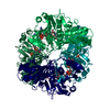 2dbvS S: Starting model for refinement |
|---|---|
| Similar structure data |


-Links
 PDBj
PDBj


- Assembly
| Deposited unit | 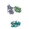
| ||||||||||
|---|---|---|---|---|---|---|---|---|---|---|---|
| 1 | 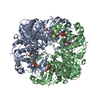
| ||||||||||
| 2 | 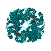
| ||||||||||
| Unit cell |
| ||||||||||
| Details | One tetramer with crystallographic 222 symmetry is generated from the monomer O by the operations -x, -y, z and -x, y, -z and x, -y, -z another similar tetramer with a crystallographic 2 symmetry is generated from the dimer O'R' by the operation 1/2-x, 1/2-y, z. |
-Components
| #1: Protein | Mass: 36012.160 Da / Num. of mol.: 3 / Source method: isolated from a natural source / Details: tetramer GAPDH 2 / Source: (natural) Spinacia oleracea (spinach) / Cellular location: CHLOROPLASTReferences: UniProt: P19866, glyceraldehyde-3-phosphate dehydrogenase (NADP+) (phosphorylating)#2: Chemical | ChemComp-SO4 / Sulfate  Mass: 96.063 Da / Num. of mol.: 6 / Source method: obtained synthetically / Formula: SO4 Mass: 96.063 Da / Num. of mol.: 6 / Source method: obtained synthetically / Formula: SO4#3: Chemical | Nicotinamide adenine dinucleotide phosphate  Mass: 745.421 Da / Num. of mol.: 3 / Source method: obtained synthetically / Formula: C21H30N7O17P3 Mass: 745.421 Da / Num. of mol.: 3 / Source method: obtained synthetically / Formula: C21H30N7O17P3#4: Water | ChemComp-HOH / | Water Mass: 18.015 Da / Num. of mol.: 212 / Source method: isolated from a natural source / Formula: H2O Mass: 18.015 Da / Num. of mol.: 212 / Source method: isolated from a natural source / Formula: H2O |
|---|
-Experimental details
-Experiment
| Experiment | Method: X-RAY DIFFRACTION / Number of used crystals: 1 |
|---|
- Sample preparation
| Crystal | Density Matthews: 3.27 Å3/Da / Density % sol: 62.36 % | ||||||||||||||||||||||||||||||||||||
|---|---|---|---|---|---|---|---|---|---|---|---|---|---|---|---|---|---|---|---|---|---|---|---|---|---|---|---|---|---|---|---|---|---|---|---|---|---|
| Crystal grow | Temperature: 298 K / Method: vapor diffusion, hanging drop / pH: 8.5 Details: Ammonium sulphate, Tris-HCl, pH 8.5, VAPOR DIFFUSION, HANGING DROP at 298K | ||||||||||||||||||||||||||||||||||||
| Crystal grow | *PLUS Temperature: 293 K / pH: 7 Details: Sabatino, P., (1999) Acta Crystallogr., Sect.D, 55, 566. | ||||||||||||||||||||||||||||||||||||
| Components of the solutions | *PLUS
|
-Data collection
| Diffraction | Mean temperature: 100 K |
|---|---|
| Diffraction source | Source: SYNCHROTRON / Site: ELETTRA  / Beamline: 5.2R / Wavelength: 1 Å / Beamline: 5.2R / Wavelength: 1 Å |
| Detector | Type: MARRESEARCH / Detector: IMAGE PLATE / Date: May 3, 1999 |
| Radiation | Monochromator: Si(111) / Protocol: SINGLE WAVELENGTH / Monochromatic (M) / Laue (L): M / Scattering type: x-ray |
| Radiation wavelength | Wavelength: 1 Å / Relative weight: 1 |
| Reflection | Resolution: 3→69 Å / Num. all: 28763 / Num. obs: 26762 / % possible obs: 93 % / Observed criterion σ(I): -3 / Redundancy: 4 % / Rmerge(I) obs: 0.13 / Rsym value: 0.132 / Net I/σ(I): 7.11 |
| Reflection shell | Resolution: 3→3.11 Å / Rmerge(I) obs: 0.499 / Num. unique all: 2308 / Rsym value: 0.437 / % possible all: 80.8 |
| Reflection | *PLUS % possible obs: 93 % |
- Processing
| Software |
| ||||||||||||||||||||||||||||||||||||||||
|---|---|---|---|---|---|---|---|---|---|---|---|---|---|---|---|---|---|---|---|---|---|---|---|---|---|---|---|---|---|---|---|---|---|---|---|---|---|---|---|---|---|
| Refinement | Method to determine structure: MOLECULAR REPLACEMENT Starting model: 2DBV Resolution: 3→49.58 Å / Rfactor Rfree error: 0.006 / Data cutoff high absF: 4360318.29 / Data cutoff low absF: 0 / Isotropic thermal model: RESTRAINED / Cross valid method: THROUGHOUT / σ(F): 0 / σ(I): 0 / Stereochemistry target values: Engh & Huber Details: Chains A and B correspond to O' and R' respectively. Arg23, Asp70 and Ala337 were deleted from the model because the electron density did not allow their positioning. Each monomer is numbered 1 to 337.
| ||||||||||||||||||||||||||||||||||||||||
| Solvent computation | Solvent model: FLAT MODEL / Bsol: 49.1735 Å2 / ksol: 0.37947 e/Å3 | ||||||||||||||||||||||||||||||||||||||||
| Displacement parameters | Biso mean: 44.1 Å2
| ||||||||||||||||||||||||||||||||||||||||
| Refine analyze |
| ||||||||||||||||||||||||||||||||||||||||
| Refinement step | Cycle: LAST / Resolution: 3→49.58 Å
| ||||||||||||||||||||||||||||||||||||||||
| Refine LS restraints |
| ||||||||||||||||||||||||||||||||||||||||
| LS refinement shell | Resolution: 3→3.19 Å / Rfactor Rfree error: 0.023 / Total num. of bins used: 6
| ||||||||||||||||||||||||||||||||||||||||
| Xplor file |
| ||||||||||||||||||||||||||||||||||||||||
| Software | *PLUS Name: CNS / Version: 1 / Classification: refinement | ||||||||||||||||||||||||||||||||||||||||
| Refinement | *PLUS Lowest resolution: 69 Å / σ(F): 0 / % reflection Rfree: 5 % | ||||||||||||||||||||||||||||||||||||||||
| Solvent computation | *PLUS | ||||||||||||||||||||||||||||||||||||||||
| Displacement parameters | *PLUS Biso mean: 44.1 Å2 | ||||||||||||||||||||||||||||||||||||||||
| Refine LS restraints | *PLUS
| ||||||||||||||||||||||||||||||||||||||||
| LS refinement shell | *PLUS Rfactor Rfree: 0.386 / % reflection Rfree: 7.4 % / Rfactor Rwork: 0.32 |
