 Movie
Movie Controller


+ Open data

- Basic information
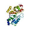
| Entry | Database: PDB / ID: 1rhs | ||||||
|---|---|---|---|---|---|---|---|
| Title | SULFUR-SUBSTITUTED RHODANESE | ||||||
 Components Components | SULFUR-SUBSTITUTED RHODANESE | ||||||
 Keywords Keywords |  TRANSFERASE / RHODANESE / SULFURTRANSFERASE TRANSFERASE / RHODANESE / SULFURTRANSFERASE | ||||||
| Function / homology |  Function and homology information Function and homology informationrRNA transport / 3-mercaptopyruvate sulfurtransferase activity / thiosulfate sulfurtransferase / thiosulfate sulfurtransferase activity / rRNA import into mitochondrion / 5S rRNA binding / mitochondrial matrix / mitochondrionSimilarity search - Function | ||||||
| Biological species |  Bos taurus (cattle) Bos taurus (cattle) | ||||||
| Method | X-RAY DIFFRACTION / Resolution: 1.36 Å | ||||||
 Authors Authors | Zanotti, G. / Gliubich, F. / Colapietro, M. / Barba, L. | ||||||
 Citation Citation | Journal: Acta Crystallogr.,Sect.D / Year: 1998 Title: Structure of sulfur-substituted rhodanese at 1.36 A resolution. Authors: Gliubich, F. / Berni, R. / Colapietro, M. / Barba, L. / Zanotti, G. #1: Journal: J.Mol.Biol. / Year: 1979Title: The Structure of Bovine Liver Rhodanese. II. The Active Site in the Sulfur-Substituted and the Sulfur-Free Enzyme Authors: Ploegman, J.H. / Drent, G. / Kalk, K.H. / Hol, W.G. | ||||||
| History |
|
- Structure visualization
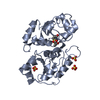
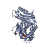
| Structure viewer | Molecule: MolmilJmol/JSmol |
|---|
- Downloads & links
-Download
| PDBx/mmCIF format | 1rhs.cif.gz | 78.9 KB | Display | PDBx/mmCIF format |
|---|---|---|---|---|
| PDB format | pdb1rhs.ent.gz | 59.4 KB | Display | PDB format |
| PDBx/mmJSON format | 1rhs.json.gz | Tree view | PDBx/mmJSON format | |
| Others |  Other downloads Other downloads |
-Validation report
| Arichive directory | https://data.pdbj.org/pub/pdb/validation_reports/rh/1rhsftp://data.pdbj.org/pub/pdb/validation_reports/rh/1rhs | HTTPS FTP |
|---|
-Related structure data
| Similar structure data |
|---|


-Links
 PDBj
PDBj- Assembly
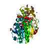
| Deposited unit | 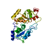
| ||||||||
|---|---|---|---|---|---|---|---|---|---|
| 1 |
| ||||||||
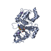
| Unit cell |
|
-Components
| #1: Protein | Mass: 33240.668 Da / Num. of mol.: 1 / Source method: isolated from a natural source / Source: (natural) Bos taurus (cattle) / Cellular location: CYTOPLASM / Organ: LIVER / References: UniProt: P00586, thiosulfate sulfurtransferase |
|---|---|
| #2: Water | ChemComp-HOH / Water Mass: 18.015 Da / Num. of mol.: 407 / Source method: isolated from a natural source / Formula: H2O Mass: 18.015 Da / Num. of mol.: 407 / Source method: isolated from a natural source / Formula: H2O |
-Experimental details
-Experiment
| Experiment | Method: X-RAY DIFFRACTION / Number of used crystals: 3 |
|---|
- Sample preparation
| Crystal | Density Matthews: 2.43 Å3/Da / Density % sol: 49 % | |||||||||||||||
|---|---|---|---|---|---|---|---|---|---|---|---|---|---|---|---|---|
| Crystal grow | pH: 7.6 / Details: pH 7.6 | |||||||||||||||
| Crystal grow | *PLUS Temperature: 20 ℃ / pH: 7.4 / Method: vapor diffusion, sitting drop | |||||||||||||||
| Components of the solutions | *PLUS
|
-Data collection
| Diffraction | Mean temperature: 100 K |
|---|---|
| Diffraction source | Wavelength: 1 |
| Detector | Type: MARRESEARCH / Detector: IMAGE PLATE AREA DETECTOR / Date: Jul 1, 1996 / Details: MIRRORS |
| Radiation | Monochromator: SI(111) / Monochromatic (M) / Laue (L): M / Scattering type: x-ray |
| Radiation wavelength | Wavelength: 1 Å / Relative weight: 1 |
| Reflection | Resolution: 1.36→25 Å / Num. obs: 56060 / % possible obs: 74 % / Observed criterion σ(I): 0 / Redundancy: 2.2 % / Rmerge(I) obs: 0.067 / Rsym value: 0.067 / Net I/σ(I): 22.6 |
| Reflection shell | Resolution: 1.3→1.4 Å / Redundancy: 1.3 % / Rmerge(I) obs: 0.33 / Mean I/σ(I) obs: 3.7 / Rsym value: 0.33 / % possible all: 45 |
| Reflection | *PLUS Num. measured all: 131477 |
| Reflection shell | *PLUS Highest resolution: 1.36 Å / Lowest resolution: 1.43 Å / % possible obs: 45 % / Num. unique obs: 10176 |
- Processing
| Software |
| |||||||||||||||||||||||||||||||||
|---|---|---|---|---|---|---|---|---|---|---|---|---|---|---|---|---|---|---|---|---|---|---|---|---|---|---|---|---|---|---|---|---|---|---|
| Refinement | Resolution: 1.36→25 Å / Num. parameters: 24750 / Num. restraintsaints: 29262 / Cross valid method: FREE R-VALUE / σ(F): 0 / Stereochemistry target values: SHELXL
| |||||||||||||||||||||||||||||||||
| Refine analyze | Num. disordered residues: 0 / Occupancy sum hydrogen: 8872 / Occupancy sum non hydrogen: 11056 | |||||||||||||||||||||||||||||||||
| Refinement step | Cycle: LAST / Resolution: 1.36→25 Å
| |||||||||||||||||||||||||||||||||
| Refine LS restraints |
| |||||||||||||||||||||||||||||||||
| Software | *PLUS Name: SHELXL-93 / Classification: refinement | |||||||||||||||||||||||||||||||||
| Refinement | *PLUS Num. reflection all: 53034 | |||||||||||||||||||||||||||||||||
| Solvent computation | *PLUS | |||||||||||||||||||||||||||||||||
| Displacement parameters | *PLUS | |||||||||||||||||||||||||||||||||
| Refine LS restraints | *PLUS
|
