 Movie
Movie Controller

[English] 日本語
 Yorodumi
- PDB-1r70: Model of human IgA2 determined by solution scattering, curve fitt... -
+ Open data

- Basic information
| Entry | Database: PDB / ID: 1r70 | ||||||
|---|---|---|---|---|---|---|---|
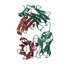
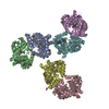
| Title | Model of human IgA2 determined by solution scattering, curve fitting and homology modelling | ||||||
 Components Components |
| ||||||
 Keywords Keywords |  IMMUNE SYSTEM / Immunology / antibody / IgA / glycoprotein / Ig fold IMMUNE SYSTEM / Immunology / antibody / IgA / glycoprotein / Ig fold | ||||||
| Biological species |  Homo sapiens (human) Homo sapiens (human) | ||||||
| Method | SOLUTION SCATTERING / SYNCHROTRON / NUCLEAR REACTOR / CONSTRAINED MODEL FIT / Resolution: 30 Å | ||||||
 Authors Authors | Furtado, P.B. / Whitty, P.W. / Robertson, A. / Eaton, J.T. / Almogren, A. / Kerr, M.A. / Woof, J.M. / Perkins, S.J. | ||||||
 Citation Citation | Journal: J.Mol.Biol. / Year: 2004 Title: Solution Structure Determination of Monomeric Human IgA2 by X-ray and Neutron Scattering, Analytical Ultracentrifugation and Constrained Modelling: A Comparison with Monomeric Human IgA1. Authors: Furtado, P.B. / Whitty, P.W. / Robertson, A. / Eaton, J.T. / Almogren, A. / Kerr, M.A. / Woof, J.M. / Perkins, S.J. | ||||||
| History |
| ||||||
| Remark 999 | SEQUENCE At the time of processing, there were no suitable sequence database references for the ...SEQUENCE At the time of processing, there were no suitable sequence database references for the proteins in this entry. |
- Structure visualization
| Structure viewer | Molecule:  MolmilJmol/JSmol MolmilJmol/JSmol |
|---|
- Downloads & links
-Download
| PDBx/mmCIF format | 1r70.cif.gz | 55.7 KB | Display | PDBx/mmCIF format |
|---|---|---|---|---|
| PDB format | pdb1r70.ent.gz | 28.9 KB | Display | PDB format |
| PDBx/mmJSON format | 1r70.json.gz | Tree view | PDBx/mmJSON format | |
| Others |  Other downloads Other downloads |
-Validation report
| Arichive directory | https://data.pdbj.org/pub/pdb/validation_reports/r7/1r70ftp://data.pdbj.org/pub/pdb/validation_reports/r7/1r70 | HTTPS FTP |
|---|
-Related structure data
| Related structure data | |
|---|---|
| Similar structure data |


-Links
 PDBj
PDBj

- Assembly
| Deposited unit | 
|
|---|---|
| 1 |
|
-Components
| #1: Antibody | Mass: 23216.770 Da / Num. of mol.: 2 Source method: isolated from a genetically manipulated source Source: (gene. exp.) Homo sapiens (human) / Gene: human alpha 2 gene (light chain) / Plasmid: pEE6.HCMV / Cell line (production host): Chinese hamster ovary (CHO) / Organ (production host): ovary / Production host:   Cricetulus griseus (Chinese hamster) / Strain (production host): K1 Cricetulus griseus (Chinese hamster) / Strain (production host): K1#2: Antibody | Mass: 49922.031 Da / Num. of mol.: 2 Source method: isolated from a genetically manipulated source Source: (gene. exp.) Homo sapiens (human) / Gene: human alpha 2 gene (heavy chain) / Plasmid: pEE6.HCMV / Cell line (production host): Chinese hamster ovary (CHO) / Organ (production host): ovary / Production host: Cricetulus griseus (Chinese hamster) / Strain (production host): K1 |
|---|
-Experimental details
-Experiment
| Experiment | Method: SOLUTION SCATTERING |
|---|
-Data collection
| Diffraction |
| ||||||||||||||||||||||||||||||||||||||||||||||||||||||||||||||||||||||||
|---|---|---|---|---|---|---|---|---|---|---|---|---|---|---|---|---|---|---|---|---|---|---|---|---|---|---|---|---|---|---|---|---|---|---|---|---|---|---|---|---|---|---|---|---|---|---|---|---|---|---|---|---|---|---|---|---|---|---|---|---|---|---|---|---|---|---|---|---|---|---|---|---|---|
| Diffraction source |
| ||||||||||||||||||||||||||||||||||||||||||||||||||||||||||||||||||||||||
| Detector |
| ||||||||||||||||||||||||||||||||||||||||||||||||||||||||||||||||||||||||
| Radiation |
| ||||||||||||||||||||||||||||||||||||||||||||||||||||||||||||||||||||||||
| Radiation wavelength | Relative weight: 1 | ||||||||||||||||||||||||||||||||||||||||||||||||||||||||||||||||||||||||
| Soln scatter | Data analysis software list: SCTPL7, GNOM / Sample pH: 7.4 / Temperature: 288 K
|


- Processing
| Software |
| |||||||||||||||||||||||||||||||||||||||||||||
|---|---|---|---|---|---|---|---|---|---|---|---|---|---|---|---|---|---|---|---|---|---|---|---|---|---|---|---|---|---|---|---|---|---|---|---|---|---|---|---|---|---|---|---|---|---|---|
| Refinement | Method to determine structure: CONSTRAINED MODEL FIT / Resolution: 30→1300 Å / Rfactor all: 0.066 / Stereochemistry target values: ENGH & HUBER | |||||||||||||||||||||||||||||||||||||||||||||
| Refinement step | Cycle: LAST / Resolution: 30→1300 Å
| |||||||||||||||||||||||||||||||||||||||||||||
| Soln scatter model | Method: CONSTRAINED SCATTERING FITTING OF HOMOLOGY MODELS Conformer selection criteria: THE MODELLED SCATTERING CURVES WERE ASSESSED BY CALCULATION OF THE RG, RSX-1 AND VALUES IN THE SAME Q RANGES USED IN THE EXPERIMENTAL GUINIER FITS. MODELS WERE THEN ...Conformer selection criteria: THE MODELLED SCATTERING CURVES WERE ASSESSED BY CALCULATION OF THE RG, RSX-1 AND VALUES IN THE SAME Q RANGES USED IN THE EXPERIMENTAL GUINIER FITS. MODELS WERE THEN RANKED USING A GOODNESS-OF-FIT R-FACTOR DEFINED BY ANALOGY WITH PROTEIN CRYSTALLOGRAPHY AND BASED ON THE EXPERIMENTAL CURVES IN THE Q RANGE EXTENDING TO 2.2 NM-1 (ESRF X-RAYS) AND 2.2 NM-1 (ISIS NEUTRONS). Details: HOMOLOGY MODELS WERE BUILT FOR THE IGA2 FAB AND FC FRAGMENTS STARTING FROM THE IGA1 MODEL (PDB ENTRY 1IGA). THE POSITIONS OF THE FAB FRAGMENTS RELATIVE TO THE FC FRAGMENT WERE DETERMINED BY ...Details: HOMOLOGY MODELS WERE BUILT FOR THE IGA2 FAB AND FC FRAGMENTS STARTING FROM THE IGA1 MODEL (PDB ENTRY 1IGA). THE POSITIONS OF THE FAB FRAGMENTS RELATIVE TO THE FC FRAGMENT WERE DETERMINED BY AN APPROACH THAT COMBINED RANDOM HINGE PEPTIDE STRUCTURES PRODUCED BY MOLECULAR DYNAMICS SIMULATIONS WITH CURVE-FITTING TO EXPERIMENTAL SOLUTION SCATTERING DATA. THE X-RAY AND NEUTRON SCATTERING CURVE I(Q) WAS CALCULATED ASSUMING A UNIFORM SCATTERING DENSITY FOR THE SPHERES USING THE DEBYE EQUATION AS ADAPTED TO SPHERES. X-RAY CURVES WERE CALCULATED FROM THE HYDRATED SPHERE MODELS WITHOUT CORRECTIONS FOR WAVELENGTH SPREAD OR BEAM DIVERGENCE, WHILE THESE CORRECTIONS WERE APPLIED FOR THE NEUTRON CURVES BUT NOW USING UNHYDRATED MODELS. A SINGLE ARRANGEMENT OF THE FAB FRAGMENTS IS PRESENTED, WHICH IS REPRESENTATIVE OF A FAMILY OF STRUCTURES THAT FIT THE SCATTERING DATA. MORE DETAILS ON THE MODELLING STRATEGY ARE CONTAINED IN THE PRIMARY REFERENCE. Num. of conformers calculated: 10000 / Num. of conformers submitted: 1 / Representative conformer: 1 / Software author list: ACCELRYS Software list: INSIGHT II, HOMOLOGY, DISCOVERY, BIOPOLYMER, DELPHI, O, SCTPL7, GNOM |
