 Movie
Movie Controller

[English] 日本語
 Yorodumi
- PDB-1r6u: Crystal structure of an active fragment of human tryptophanyl-tRN... -
+ Open data

- Basic information
| Entry | Database: PDB / ID: 1r6u | ||||||
|---|---|---|---|---|---|---|---|
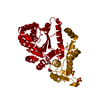
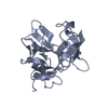
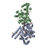
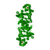
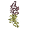
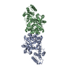
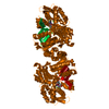
| Title | Crystal structure of an active fragment of human tryptophanyl-tRNA synthetase with cytokine activity | ||||||
 Components Components | Tryptophanyl-tRNA synthetase | ||||||
 Keywords Keywords |  LIGASE / Class Ic tRNA synthetase / Rossmann fold catalytic domain / anticodon recognition domain / bound Trp-AMP LIGASE / Class Ic tRNA synthetase / Rossmann fold catalytic domain / anticodon recognition domain / bound Trp-AMP | ||||||
| Function / homology |  Function and homology informationtryptophan-tRNA ligase / tryptophan-tRNA ligase activity / tryptophanyl-tRNA aminoacylation / kinase inhibitor activity / Cytosolic tRNA aminoacylation / regulation of angiogenesis / positive regulation of protein-containing complex assembly / negative regulation of protein kinase activity / angiogenesis / translation ...tryptophan-tRNA ligase / tryptophan-tRNA ligase activity / tryptophanyl-tRNA aminoacylation / kinase inhibitor activity / Cytosolic tRNA aminoacylation / regulation of angiogenesis / positive regulation of protein-containing complex assembly / negative regulation of protein kinase activity / angiogenesis / translation / protein domain specific binding / negative regulation of cell population proliferation / positive regulation of gene expression / protein kinase binding / protein homodimerization activity / protein-containing complex / extracellular exosome / ATP binding / nucleus / cytosol / cytoplasm Function and homology informationtryptophan-tRNA ligase / tryptophan-tRNA ligase activity / tryptophanyl-tRNA aminoacylation / kinase inhibitor activity / Cytosolic tRNA aminoacylation / regulation of angiogenesis / positive regulation of protein-containing complex assembly / negative regulation of protein kinase activity / angiogenesis / translation ...tryptophan-tRNA ligase / tryptophan-tRNA ligase activity / tryptophanyl-tRNA aminoacylation / kinase inhibitor activity / Cytosolic tRNA aminoacylation / regulation of angiogenesis / positive regulation of protein-containing complex assembly / negative regulation of protein kinase activity / angiogenesis / translation / protein domain specific binding / negative regulation of cell population proliferation / positive regulation of gene expression / protein kinase binding / protein homodimerization activity / protein-containing complex / extracellular exosome / ATP binding / nucleus / cytosol / cytoplasmSimilarity search - Function | ||||||
| Biological species |  Homo sapiens (human) Homo sapiens (human) | ||||||
| Method | X-RAY DIFFRACTION / SYNCHROTRON / MOLECULAR REPLACEMENT / Resolution: 2 Å | ||||||
 Authors Authors | Yang, X.-L. / Otero, F.J. / Skene, R.J. / McRee, D.E. / Ribas de Pouplana, L. / Schimmel, P. | ||||||
 Citation Citation | Journal: Structure / Year: 2007 Title: Functional and crystal structure analysis of active site adaptations of a potent anti-angiogenic human tRNA synthetase Authors: Yang, X.-L. / Guo, M. / Kapoor, M. / Ewalt, K.L. / Otero, F.J. / Skene, R.J. / McRee, D.E. / Schimmel, P. | ||||||
| History |
|
- Structure visualization
| Structure viewer | Molecule: MolmilJmol/JSmol |
|---|
- Downloads & links
-Download
| PDBx/mmCIF format | 1r6u.cif.gz | 162.7 KB | Display | PDBx/mmCIF format |
|---|---|---|---|---|
| PDB format | pdb1r6u.ent.gz | 134 KB | Display | PDB format |
| PDBx/mmJSON format | 1r6u.json.gz | Tree view | PDBx/mmJSON format | |
| Others |  Other downloads Other downloads |
-Validation report
| Arichive directory | https://data.pdbj.org/pub/pdb/validation_reports/r6/1r6uftp://data.pdbj.org/pub/pdb/validation_reports/r6/1r6u | HTTPS FTP |
|---|
-Related structure data
| Related structure data | |
|---|---|
| Similar structure data |


-Links
 PDBj
PDBj
- Assembly
| Deposited unit | 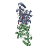
| ||||||||
|---|---|---|---|---|---|---|---|---|---|
| 1 |
| ||||||||
| Unit cell |
| ||||||||
| Components on special symmetry positions |
|
-Components
| #1: Protein | Mass: 50158.098 Da / Num. of mol.: 2 / Mutation: S213G, Y214D Source method: isolated from a genetically manipulated source Source: (gene. exp.) Homo sapiens (human) / Gene: WARS / Plasmid: pET20b+ / Production host:  Escherichia coli (E. coli) / Strain (production host): B834(DE3) / References: UniProt: P23381, tryptophan-tRNA ligase Escherichia coli (E. coli) / Strain (production host): B834(DE3) / References: UniProt: P23381, tryptophan-tRNA ligase#2: Chemical | ChemComp-TYM / |   Mass: 533.431 Da / Num. of mol.: 1 / Source method: obtained synthetically / Formula: C21H24N7O8P Mass: 533.431 Da / Num. of mol.: 1 / Source method: obtained synthetically / Formula: C21H24N7O8P#3: Chemical | Glycerol  Mass: 92.094 Da / Num. of mol.: 2 / Source method: obtained synthetically / Formula: C3H8O3 Mass: 92.094 Da / Num. of mol.: 2 / Source method: obtained synthetically / Formula: C3H8O3#4: Water | ChemComp-HOH / | Water Mass: 18.015 Da / Num. of mol.: 100 / Source method: isolated from a natural source / Formula: H2O Mass: 18.015 Da / Num. of mol.: 100 / Source method: isolated from a natural source / Formula: H2O |
|---|
-Experimental details
-Experiment
| Experiment | Method: X-RAY DIFFRACTION / Number of used crystals: 1 |
|---|
- Sample preparation
| Crystal | Density Matthews: 2.41 Å3/Da / Density % sol: 48.98 % | |||||||||||||||||||||||||||||||||||||||||||||||||
|---|---|---|---|---|---|---|---|---|---|---|---|---|---|---|---|---|---|---|---|---|---|---|---|---|---|---|---|---|---|---|---|---|---|---|---|---|---|---|---|---|---|---|---|---|---|---|---|---|---|---|
| Crystal grow | Temperature: 279 K / Method: vapor diffusion, sitting drop / pH: 7.02 Details: PEG MME 550, Hepes, pH 7.02, VAPOR DIFFUSION, SITTING DROP, temperature 279K | |||||||||||||||||||||||||||||||||||||||||||||||||
| Crystal grow | *PLUS Temperature: 4 ℃ / pH: 7.5 / Method: vapor diffusion, sitting drop | |||||||||||||||||||||||||||||||||||||||||||||||||
| Components of the solutions | *PLUS
|
-Data collection
| Diffraction | Mean temperature: 100 K |
|---|---|
| Diffraction source | Source: SYNCHROTRON / Site: SSRL  / Beamline: BL9-2 / Wavelength: 0.91162 Å / Beamline: BL9-2 / Wavelength: 0.91162 Å |
| Detector | Type: ADSC QUANTUM 315 / Detector: CCD / Date: Dec 22, 2002 |
| Radiation | Monochromator: double crystal monochromator / Protocol: SINGLE WAVELENGTH / Monochromatic (M) / Laue (L): M / Scattering type: x-ray |
| Radiation wavelength | Wavelength: 0.91162 Å / Relative weight: 1 |
| Reflection | Resolution: 2→30 Å / Num. all: 127115 / Num. obs: 126480 / % possible obs: 99.5 % / Redundancy: 7.3 % / Rmerge(I) obs: 0.038 / Rsym value: 0.038 / Net I/σ(I): 34.7 |
| Reflection shell | Resolution: 2→2.07 Å / % possible all: 99.5 |
| Reflection | *PLUS Num. obs: 64590 |
| Reflection shell | *PLUS % possible obs: 99.6 % / Rmerge(I) obs: 0.587 / Mean I/σ(I) obs: 2 |
- Processing
| Software |
| ||||||||||||||||||||
|---|---|---|---|---|---|---|---|---|---|---|---|---|---|---|---|---|---|---|---|---|---|
| Refinement | Method to determine structure: MOLECULAR REPLACEMENT Starting model: model derived from the structure of the full-length human TrpRS Resolution: 2→30 Å / Stereochemistry target values: Engh & Huber
| ||||||||||||||||||||
| Solvent computation | Bsol: 54.3854 Å2 / ksol: 0.360966 e/Å3 | ||||||||||||||||||||
| Displacement parameters |
| ||||||||||||||||||||
| Refinement step | Cycle: LAST / Resolution: 2→30 Å
| ||||||||||||||||||||
| Refine LS restraints |
| ||||||||||||||||||||
| Xplor file |
| ||||||||||||||||||||
| Refinement | *PLUS Lowest resolution: 20 Å / % reflection Rfree: 5 % | ||||||||||||||||||||
| Solvent computation | *PLUS | ||||||||||||||||||||
| Displacement parameters | *PLUS | ||||||||||||||||||||
| Refine LS restraints | *PLUS
|
