 Movie
Movie Controller


+ Open data

- Basic information
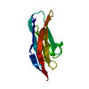
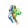
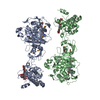
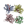
| Entry | Database: PDB / ID: 1qz1 | ||||||
|---|---|---|---|---|---|---|---|
| Title | Crystal Structure of the Ig 1-2-3 fragment of NCAM | ||||||
 Components Components | Neural cell adhesion molecule 1, 140 kDa isoform | ||||||
 Keywords Keywords | CELL ADHESION / IG MODULES / NCAM | ||||||
| Function / homology |  Function and homology information Function and homology informationNCAM1 interactions / regulation of exocyst assembly / regulation of semaphorin-plexin signaling pathway / calcium-independent cell-cell adhesion via plasma membrane cell-adhesion molecules / cellular response to molecule of bacterial origin / peripheral nervous system axon regeneration / LRR domain binding / homotypic cell-cell adhesion / NCAM signaling for neurite out-growth / Signal transduction by L1 ...NCAM1 interactions / regulation of exocyst assembly / regulation of semaphorin-plexin signaling pathway / calcium-independent cell-cell adhesion via plasma membrane cell-adhesion molecules / cellular response to molecule of bacterial origin / peripheral nervous system axon regeneration / LRR domain binding / homotypic cell-cell adhesion / NCAM signaling for neurite out-growth / Signal transduction by L1 / cellular response to inorganic substance / thalamus development / fibroblast growth factor receptor binding / commissural neuron axon guidance / axonal fasciculation / negative regulation of programmed cell death / RAF/MAP kinase cascade / response to inorganic substance / neuron development / epithelial to mesenchymal transition / multicellular organismal response to stress / phosphatase binding / animal organ regeneration / positive regulation of calcium-mediated signaling / positive regulation of cardiac muscle cell proliferation / cytoskeletal protein binding / response to activity / response to cocaine / calcium-mediated signaling / response to lead ion / modulation of chemical synaptic transmission / Schaffer collateral - CA1 synapse / neuron projection development / cell-cell junction / presynaptic membrane / heparin binding / growth cone / postsynaptic membrane / learning or memory / cell surface receptor signaling pathway / cell adhesion / response to xenobiotic stimulus / axon / external side of plasma membrane / neuronal cell body / glutamatergic synapse / cell surface / plasma membraneSimilarity search - Function | ||||||
| Biological species |  Rattus norvegicus (Norway rat) Rattus norvegicus (Norway rat) | ||||||
| Method | X-RAY DIFFRACTION / SYNCHROTRON / MOLECULAR REPLACEMENT / Resolution: 2 Å | ||||||
 Authors Authors | Soroka, V. / Kolkova, K. / Kastrup, J.S. / Diederichs, K. / Breed, J. / Kiselyov, V.V. / Poulsen, F.M. / Larsen, I.K. / Welte, W. / Berezin, V. ...Soroka, V. / Kolkova, K. / Kastrup, J.S. / Diederichs, K. / Breed, J. / Kiselyov, V.V. / Poulsen, F.M. / Larsen, I.K. / Welte, W. / Berezin, V. / Bock, E. / Kasper, C. | ||||||
 Citation Citation | Journal: Structure / Year: 2003 Title: Structure and interactions of NCAM Ig1-2-3 suggest a novel zipper mechanism for homophilic adhesion Authors: Soroka, V. / Kolkova, K. / Kastrup, J.S. / Diederichs, K. / Breed, J. / Kiselyov, V.V. / Poulsen, F.M. / Larsen, I.K. / Welte, W. / Berezin, V. / Bock, E. / Kasper, C. #1: Journal: Nat.Struct.Biol. / Year: 2000Title: STRUCTURAL BASIS OF CELL-CELL ADHESION BY NCAM Authors: Kasper, C. / Rasmussen, H. / Kastrup, J.S. / Ikemizu, S. / Jones, E.Y. / Berezin, V. / Bock, E. / Larsen, I.K. #2: Journal: Acta Crystallogr.,Sect.D / Year: 1999Title: Expression, crystallization and preliminary X-ray analysis of the two amino-terminal Ig domains of the neural cell adhesion molecule (NCAM) Authors: Kasper, C. / Rasmussen, H. / Berezin, V. / Bock, E. / Larsen, I.K. | ||||||
| History |
| ||||||
| Remark 999 | SEQUENCE Residues -2, 239 and 240 were not visible in the electron density. |
- Structure visualization
| Structure viewer | Molecule: MolmilJmol/JSmol |
|---|
- Downloads & links
-Download
| PDBx/mmCIF format | 1qz1.cif.gz | 77.4 KB | Display | PDBx/mmCIF format |
|---|---|---|---|---|
| PDB format | pdb1qz1.ent.gz | 56.4 KB | Display | PDB format |
| PDBx/mmJSON format | 1qz1.json.gz | Tree view | PDBx/mmJSON format | |
| Others |  Other downloads Other downloads |
-Validation report
| Arichive directory | https://data.pdbj.org/pub/pdb/validation_reports/qz/1qz1ftp://data.pdbj.org/pub/pdb/validation_reports/qz/1qz1 | HTTPS FTP |
|---|
-Related structure data
| Related structure data |  1epfS S: Starting model for refinement |
|---|---|
| Similar structure data |

-Links
 PDBj
PDBj


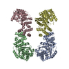
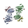
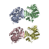
- Assembly
| Deposited unit | 
| ||||||||
|---|---|---|---|---|---|---|---|---|---|
| 1 | 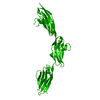
| ||||||||
| Unit cell |
| ||||||||
| Details | The biological assembly is a dimer. The second part of the biological assembly is generated by the rotation (-x, y, -z) and the translation (0.5, 0, 0) |
-Components
| #1: Protein | / N-CAM 140 / NCAM-140 Mass: 32407.225 Da / Num. of mol.: 1 / Fragment: IG MODULES 1-2-3 Source method: isolated from a genetically manipulated source Source: (gene. exp.) Rattus norvegicus (Norway rat) / Gene: NCAM1 / Plasmid: pHIL-S1 / Production host:  Pichia pastoris (fungus) / Strain (production host): GS-115 / References: UniProt: P13596 Pichia pastoris (fungus) / Strain (production host): GS-115 / References: UniProt: P13596 |
|---|---|
| #2: Water | ChemComp-HOH / Water Mass: 18.015 Da / Num. of mol.: 265 / Source method: isolated from a natural source / Formula: H2O Mass: 18.015 Da / Num. of mol.: 265 / Source method: isolated from a natural source / Formula: H2O |
-Experimental details
-Experiment
| Experiment | Method: X-RAY DIFFRACTION / Number of used crystals: 1 |
|---|
- Sample preparation
| Crystal | Density Matthews: 3.19 Å3/Da / Density % sol: 61.45 % | |||||||||||||||||||||||||||||||||||||||||||||||||
|---|---|---|---|---|---|---|---|---|---|---|---|---|---|---|---|---|---|---|---|---|---|---|---|---|---|---|---|---|---|---|---|---|---|---|---|---|---|---|---|---|---|---|---|---|---|---|---|---|---|---|
| Crystal grow | Temperature: 293 K / Method: vapor diffusion, hanging drop / pH: 5.2 Details: 14-17% PEG 4000, 450 mM Li sulfate, 100 mM Na acetate, pH 5.2, VAPOR DIFFUSION, HANGING DROP, temperature 293K | |||||||||||||||||||||||||||||||||||||||||||||||||
| Crystal grow | *PLUS Temperature: 18 ℃ / pH: 7.4 / Method: vapor diffusion, hanging drop | |||||||||||||||||||||||||||||||||||||||||||||||||
| Components of the solutions | *PLUS
|
-Data collection
| Diffraction |
| |||||||||||||||||||||
|---|---|---|---|---|---|---|---|---|---|---|---|---|---|---|---|---|---|---|---|---|---|---|
| Diffraction source |
| |||||||||||||||||||||
| Detector |
| |||||||||||||||||||||
| Radiation | Protocol: SINGLE WAVELENGTH / Monochromatic (M) / Laue (L): M / Scattering type: x-ray | |||||||||||||||||||||
| Radiation wavelength |
| |||||||||||||||||||||
| Reflection | Resolution: 2→50 Å / Num. all: 27881 / Num. obs: 27881 / % possible obs: 99.2 % / Observed criterion σ(F): 0 / Observed criterion σ(I): 0 / Redundancy: 5.9 % / Biso Wilson estimate: 42 Å2 / Rmerge(I) obs: 0.039 / Rsym value: 0.039 / Net I/σ(I): 19.6 | |||||||||||||||||||||
| Reflection shell | Resolution: 2→2.07 Å / Redundancy: 3.8 % / Rmerge(I) obs: 0.209 / Mean I/σ(I) obs: 1.4 / Num. unique all: 2756 / Rsym value: 0.209 / % possible all: 99.4 | |||||||||||||||||||||
| Reflection | *PLUS Lowest resolution: 50 Å / Num. measured all: 164206 | |||||||||||||||||||||
| Reflection shell | *PLUS % possible obs: 99.4 % |

- Processing
| Software |
| |||||||||||||||||||||||||
|---|---|---|---|---|---|---|---|---|---|---|---|---|---|---|---|---|---|---|---|---|---|---|---|---|---|---|
| Refinement | Method to determine structure: MOLECULAR REPLACEMENT Starting model: PDB ENTRY 1EPF Resolution: 2→48.64 Å / Isotropic thermal model: anisotropic / Cross valid method: THROUGHOUT / σ(F): 0 / σ(I): 0 / Stereochemistry target values: Engh & Huber Details: Residues 241-242 were not located in the electron density map
| |||||||||||||||||||||||||
| Displacement parameters | Biso mean: 60.6 Å2
| |||||||||||||||||||||||||
| Refine analyze |
| |||||||||||||||||||||||||
| Refinement step | Cycle: LAST / Resolution: 2→48.64 Å
| |||||||||||||||||||||||||
| Refine LS restraints |
| |||||||||||||||||||||||||
| LS refinement shell | Resolution: 2→2.13 Å / Rfactor Rfree error: 0.036
| |||||||||||||||||||||||||
| Refinement | *PLUS % reflection Rfree: 3 % | |||||||||||||||||||||||||
| Solvent computation | *PLUS | |||||||||||||||||||||||||
| Displacement parameters | *PLUS | |||||||||||||||||||||||||
| Refine LS restraints | *PLUS
|
