[English] 日本語
Yorodumi
- PDB-6e7k: Structure of the lipoprotein lipase GPIHBP1 complex that mediates... -

+
Open data


ID or keywords:

Loading...

-
Basic information

Entry
Database: PDB / ID: 6e7k
TitleStructure of the lipoprotein lipase GPIHBP1 complex that mediates plasma triglyceride hydrolysis
Components
  • Glycosylphosphatidylinositol-anchored high density lipoprotein-binding protein 1
  • Lipoprotein lipase
KeywordsHYDROLASE / hydrolase-cofactor complex / lipid degradation
Function / homology
Function and homology information


chylomicron binding / positive regulation of chylomicron remnant clearance / lipoprotein lipase / lipoprotein lipase activity / positive regulation of sequestering of triglyceride / low-density lipoprotein particle mediated signaling / chylomicron remodeling / response to heparin / lipase binding / positive regulation of cholesterol storage ...chylomicron binding / positive regulation of chylomicron remnant clearance / lipoprotein lipase / lipoprotein lipase activity / positive regulation of sequestering of triglyceride / low-density lipoprotein particle mediated signaling / chylomicron remodeling / response to heparin / lipase binding / positive regulation of cholesterol storage / lipoprotein lipase activator activity / phospholipase A1 / phosphatidylserine 1-acylhydrolase activity / 1-acyl-2-lysophosphatidylserine acylhydrolase activity / phospholipase A1 activity / phospholipase activity / triglyceride catabolic process / Assembly of active LPL and LIPC lipase complexes / Post-translational modification: synthesis of GPI-anchored proteins / positive regulation of lipoprotein lipase activity / protein transporter activity / very-low-density lipoprotein particle remodeling / positive regulation of macrophage derived foam cell differentiation / Chylomicron remodeling / positive regulation of lipid storage / chylomicron / acetylcholine receptor inhibitor activity / cellular response to nutrient / very-low-density lipoprotein particle / transcytosis / protein import / triglyceride lipase activity / positive regulation of chemokine (C-X-C motif) ligand 2 production / protein localization to cell surface / heparan sulfate proteoglycan binding / triglyceride homeostasis / cellular response to fatty acid / triglyceride metabolic process / lipoprotein particle binding / apolipoprotein binding / catalytic complex / positive regulation of fat cell differentiation / phospholipid metabolic process / response to glucose / protein-membrane adaptor activity / Retinoid metabolism and transport / positive regulation of chemokine production / positive regulation of adipose tissue development / fatty acid metabolic process / positive regulation of interleukin-1 beta production / cholesterol homeostasis / response to bacterium / intracellular protein transport / Transcriptional regulation of white adipocyte differentiation / positive regulation of inflammatory response / fatty acid biosynthetic process / positive regulation of interleukin-6 production / positive regulation of tumor necrosis factor production / heparin binding / basolateral plasma membrane / protein stabilization / apical plasma membrane / external side of plasma membrane / signaling receptor binding / lipid binding / calcium ion binding / cell surface / protein homodimerization activity / extracellular space / extracellular region / plasma membrane
Similarity search - Function
Lipoprotein lipase / Lipase, LIPH-type / Lipase, N-terminal / Triacylglycerol lipase family / Lipase / Lipase / u-PAR/Ly-6 domain / Ly-6 antigen/uPA receptor-like / Lipoxygenase homology 2 (beta barrel) domain / PLAT/LH2 domain ...Lipoprotein lipase / Lipase, LIPH-type / Lipase, N-terminal / Triacylglycerol lipase family / Lipase / Lipase / u-PAR/Ly-6 domain / Ly-6 antigen/uPA receptor-like / Lipoxygenase homology 2 (beta barrel) domain / PLAT/LH2 domain / PLAT/LH2 domain superfamily / PLAT/LH2 domain / PLAT domain profile. / Lipases, serine active site. / Snake toxin-like superfamily / Alpha/Beta hydrolase fold
Similarity search - Domain/homology
Lipoprotein lipase / Glycosylphosphatidylinositol-anchored high density lipoprotein-binding protein 1
Similarity search - Component
Biological speciesHomo sapiens (human)
MethodX-RAY DIFFRACTION / SYNCHROTRON / MOLECULAR REPLACEMENT / Resolution: 2.8 Å
AuthorsBirrane, G. / Meiyappan, M.
Citation
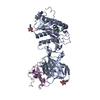
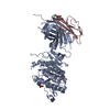
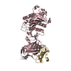
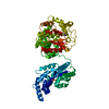
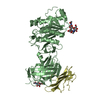
Journal: Proc Natl Acad Sci U S A / Year: 2019
Title: Structure of the lipoprotein lipase-GPIHBP1 complex that mediates plasma triglyceride hydrolysis.
Authors: Gabriel Birrane / Anne P Beigneux / Brian Dwyer / Bettina Strack-Logue / Kristian Kølby Kristensen / Omar L Francone / Loren G Fong / Haydyn D T Mertens / Clark Q Pan / Michael Ploug / ...Authors: Gabriel Birrane / Anne P Beigneux / Brian Dwyer / Bettina Strack-Logue / Kristian Kølby Kristensen / Omar L Francone / Loren G Fong / Haydyn D T Mertens / Clark Q Pan / Michael Ploug / Stephen G Young / Muthuraman Meiyappan /
Abstract: Lipoprotein lipase (LPL) is responsible for the intravascular processing of triglyceride-rich lipoproteins. The LPL within capillaries is bound to GPIHBP1, an endothelial cell protein with a three- ...Lipoprotein lipase (LPL) is responsible for the intravascular processing of triglyceride-rich lipoproteins. The LPL within capillaries is bound to GPIHBP1, an endothelial cell protein with a three-fingered LU domain and an N-terminal intrinsically disordered acidic domain. Loss-of-function mutations in or cause severe hypertriglyceridemia (chylomicronemia), but structures for LPL and GPIHBP1 have remained elusive. Inspired by our recent discovery that GPIHBP1's acidic domain preserves LPL structure and activity, we crystallized an LPL-GPIHBP1 complex and solved its structure. GPIHBP1's LU domain binds to LPL's C-terminal domain, largely by hydrophobic interactions. Analysis of electrostatic surfaces revealed that LPL contains a large basic patch spanning its N- and C-terminal domains. GPIHBP1's acidic domain was not defined in the electron density map but was positioned to interact with LPL's large basic patch, providing a likely explanation for how GPIHBP1 stabilizes LPL. The LPL-GPIHBP1 structure provides insights into mutations causing chylomicronemia.
History
DepositionJul 26, 2018Deposition site: RCSB / Processing site: RCSB
Revision 1.0Dec 19, 2018Provider: repository / Type: Initial release
Revision 1.1Jan 2, 2019Group: Data collection / Database references / Category: citation / citation_author
Item: _citation.journal_abbrev / _citation.pdbx_database_id_PubMed ..._citation.journal_abbrev / _citation.pdbx_database_id_PubMed / _citation.title / _citation_author.identifier_ORCID / _citation_author.name
Revision 1.2Feb 13, 2019Group: Data collection / Database references / Category: citation
Item: _citation.journal_volume / _citation.page_first ..._citation.journal_volume / _citation.page_first / _citation.page_last / _citation.year
Revision 2.0Jul 29, 2020Group: Atomic model / Data collection ...Atomic model / Data collection / Derived calculations / Structure summary
Category: atom_site / chem_comp ...atom_site / chem_comp / entity / pdbx_branch_scheme / pdbx_chem_comp_identifier / pdbx_entity_branch / pdbx_entity_branch_descriptor / pdbx_entity_branch_link / pdbx_entity_branch_list / pdbx_entity_nonpoly / pdbx_nonpoly_scheme / pdbx_struct_assembly_gen / pdbx_struct_conn_angle / struct_asym / struct_conn / struct_site / struct_site_gen
Item: _atom_site.B_iso_or_equiv / _atom_site.Cartn_x ..._atom_site.B_iso_or_equiv / _atom_site.Cartn_x / _atom_site.Cartn_y / _atom_site.Cartn_z / _atom_site.auth_asym_id / _atom_site.auth_atom_id / _atom_site.auth_comp_id / _atom_site.auth_seq_id / _atom_site.label_asym_id / _atom_site.label_atom_id / _atom_site.label_comp_id / _atom_site.label_entity_id / _atom_site.type_symbol / _chem_comp.name / _chem_comp.type / _pdbx_struct_assembly_gen.asym_id_list / _pdbx_struct_conn_angle.ptnr1_auth_seq_id / _pdbx_struct_conn_angle.ptnr1_label_asym_id / _pdbx_struct_conn_angle.ptnr2_label_asym_id / _pdbx_struct_conn_angle.ptnr3_auth_seq_id / _pdbx_struct_conn_angle.ptnr3_label_asym_id / _pdbx_struct_conn_angle.value / _struct_conn.conn_type_id / _struct_conn.id / _struct_conn.pdbx_dist_value / _struct_conn.pdbx_leaving_atom_flag / _struct_conn.pdbx_role / _struct_conn.ptnr1_auth_asym_id / _struct_conn.ptnr1_auth_comp_id / _struct_conn.ptnr1_auth_seq_id / _struct_conn.ptnr1_label_asym_id / _struct_conn.ptnr1_label_atom_id / _struct_conn.ptnr1_label_comp_id / _struct_conn.ptnr1_label_seq_id / _struct_conn.ptnr2_auth_asym_id / _struct_conn.ptnr2_auth_comp_id / _struct_conn.ptnr2_auth_seq_id / _struct_conn.ptnr2_label_asym_id / _struct_conn.ptnr2_label_atom_id / _struct_conn.ptnr2_label_comp_id
Description: Carbohydrate remediation / Provider: repository / Type: Remediation
Revision 2.1Oct 11, 2023Group: Data collection / Database references ...Data collection / Database references / Derived calculations / Refinement description / Structure summary
Category: chem_comp / chem_comp_atom ...chem_comp / chem_comp_atom / chem_comp_bond / database_2 / pdbx_initial_refinement_model / struct_conn / struct_ncs_dom_lim
Item: _chem_comp.pdbx_synonyms / _database_2.pdbx_DOI ..._chem_comp.pdbx_synonyms / _database_2.pdbx_DOI / _database_2.pdbx_database_accession / _struct_conn.pdbx_leaving_atom_flag / _struct_ncs_dom_lim.beg_auth_comp_id / _struct_ncs_dom_lim.beg_label_asym_id / _struct_ncs_dom_lim.beg_label_comp_id / _struct_ncs_dom_lim.beg_label_seq_id / _struct_ncs_dom_lim.end_auth_comp_id / _struct_ncs_dom_lim.end_label_asym_id / _struct_ncs_dom_lim.end_label_comp_id / _struct_ncs_dom_lim.end_label_seq_id

-
Structure visualization

Structure viewerMolecule:
MolmilJmol/JSmol

Downloads & links

-
Assembly

Deposited unit
A: Lipoprotein lipase
B: Lipoprotein lipase
C: Glycosylphosphatidylinositol-anchored high density lipoprotein-binding protein 1
D: Glycosylphosphatidylinositol-anchored high density lipoprotein-binding protein 1
hetero molecules


Theoretical massNumber of molelcules
Total (without water)132,62211
Polymers130,2104
Non-polymers2,4127
Water28816
1
A: Lipoprotein lipase
C: Glycosylphosphatidylinositol-anchored high density lipoprotein-binding protein 1
hetero molecules


Theoretical massNumber of molelcules
Total (without water)66,6856
Polymers65,1052
Non-polymers1,5814
Water362
TypeNameSymmetry operationNumber
identity operation1_555x,y,z1
2
B: Lipoprotein lipase
D: Glycosylphosphatidylinositol-anchored high density lipoprotein-binding protein 1
hetero molecules


Theoretical massNumber of molelcules
Total (without water)65,9375
Polymers65,1052
Non-polymers8323
Water362
TypeNameSymmetry operationNumber
identity operation1_555x,y,z1
Unit cell
Length a, b, c (Å)101.948, 153.206, 95.783
Angle α, β, γ (deg.)90.00, 90.00, 90.00
Int Tables number18
Space group name H-MP21212
Noncrystallographic symmetry (NCS)NCS domain:
IDEns-IDDetails
11A
21B
12C
22D

NCS domain segments:

Component-ID: 0 / Refine code: 0

Dom-IDEns-IDBeg auth comp-IDBeg label comp-IDEnd auth comp-IDEnd label comp-IDAuth asym-IDLabel asym-IDAuth seq-IDLabel seq-ID
11ASPASPSERSERAA33 - 4696 - 442
21ASPASPSERSERBB33 - 4696 - 442
12LEULEUTRPTRPCC62 - 14142 - 121
22LEULEUTRPTRPDD62 - 14142 - 121

NCS ensembles :
ID
1
2

-
Components

-
Protein , 2 types, 4 molecules ABCD

#1: Protein Lipoprotein lipase / / LPL


Mass: 50379.016 Da / Num. of mol.: 2 / Fragment: UNP residues 28-475 / Mutation: R324A
Source method: isolated from a genetically manipulated source
Source: (gene. exp.) Homo sapiens (human) / Gene: LPL, LIPD / Plasmid: pZ804N / Cell line (production host): HT1080 / Production host: Homo sapiens (human) / Tissue (production host): Connective tissue / References: UniProt: P06858, lipoprotein lipase
#2: Protein Glycosylphosphatidylinositol-anchored high density lipoprotein-binding protein 1 / GPIHBP1 / GPI-anchored HDL-binding protein 1 / High density lipoprotein-binding protein 1


Mass: 14725.788 Da / Num. of mol.: 2 / Fragment: UNP residues 21-151
Source method: isolated from a genetically manipulated source
Source: (gene. exp.) Homo sapiens (human) / Gene: GPIHBP1, HBP1 / Plasmid: pFastbac-1 / Cell line (production host): sf9 / Production host: Spodoptera frugiperda (fall armyworm) / References: UniProt: Q8IV16

-
Sugars , 4 types, 5 molecules

#3: Polysaccharide beta-D-mannopyranose-(1-4)-2-acetamido-2-deoxy-beta-D-glucopyranose-(1-4)-2-acetamido-2-deoxy-beta- ...beta-D-mannopyranose-(1-4)-2-acetamido-2-deoxy-beta-D-glucopyranose-(1-4)-2-acetamido-2-deoxy-beta-D-glucopyranose


Type: oligosaccharide / Mass: 586.542 Da / Num. of mol.: 1 / Source method: isolated from a natural source
DescriptorTypeProgram
DManpb1-4DGlcpNAcb1-4DGlcpNAcb1-Glycam Condensed SequenceGMML 1.0
WURCS=2.0/2,3,2/[a2122h-1b_1-5_2*NCC/3=O][a1122h-1b_1-5]/1-1-2/a4-b1_b4-c1WURCSPDB2Glycan 1.1.0
[]{[(4+1)][b-D-GlcpNAc]{[(4+1)][b-D-GlcpNAc]{[(4+1)][b-D-Manp]{}}}}LINUCSPDB-CARE
#4: Polysaccharide beta-D-mannopyranose-(1-4)-2-acetamido-2-deoxy-beta-D-glucopyranose-(1-4)-[alpha-L-fucopyranose-(1- ...beta-D-mannopyranose-(1-4)-2-acetamido-2-deoxy-beta-D-glucopyranose-(1-4)-[alpha-L-fucopyranose-(1-6)]2-acetamido-2-deoxy-beta-D-glucopyranose


Type: oligosaccharide / Mass: 732.682 Da / Num. of mol.: 1 / Source method: isolated from a natural source
DescriptorTypeProgram
DManpb1-4DGlcpNAcb1-4[LFucpa1-6]DGlcpNAcb1-Glycam Condensed SequenceGMML 1.0
WURCS=2.0/3,4,3/[a2122h-1b_1-5_2*NCC/3=O][a1122h-1b_1-5][a1221m-1a_1-5]/1-1-2-3/a4-b1_a6-d1_b4-c1WURCSPDB2Glycan 1.1.0
[]{[(4+1)][b-D-GlcpNAc]{[(4+1)][b-D-GlcpNAc]{[(4+1)][b-D-Manp]{}}[(6+1)][a-L-Fucp]{}}}LINUCSPDB-CARE
#5: Polysaccharide 2-acetamido-2-deoxy-beta-D-glucopyranose-(1-4)-[alpha-L-fucopyranose-(1-6)]2-acetamido-2-deoxy-beta- ...2-acetamido-2-deoxy-beta-D-glucopyranose-(1-4)-[alpha-L-fucopyranose-(1-6)]2-acetamido-2-deoxy-beta-D-glucopyranose


Type: oligosaccharide / Mass: 570.542 Da / Num. of mol.: 1 / Source method: isolated from a natural source
DescriptorTypeProgram
DGlcpNAcb1-4[LFucpa1-6]DGlcpNAcb1-Glycam Condensed SequenceGMML 1.0
WURCS=2.0/2,3,2/[a2122h-1b_1-5_2*NCC/3=O][a1221m-1a_1-5]/1-1-2/a4-b1_a6-c1WURCSPDB2Glycan 1.1.0
[]{[(4+1)][b-D-GlcpNAc]{[(4+1)][b-D-GlcpNAc]{}[(6+1)][a-L-Fucp]{}}}LINUCSPDB-CARE
#7: Sugar ChemComp-NAG / 2-acetamido-2-deoxy-beta-D-glucopyranose / N-acetyl-beta-D-glucosamine / 2-acetamido-2-deoxy-beta-D-glucose / 2-acetamido-2-deoxy-D-glucose / 2-acetamido-2-deoxy-glucose / N-ACETYL-D-GLUCOSAMINE / N-Acetylglucosamine


Type: D-saccharide, beta linking / Mass: 221.208 Da / Num. of mol.: 2 / Source method: isolated from a natural source / Formula: C8H15NO6
IdentifierTypeProgram
DGlcpNAcbCONDENSED IUPAC CARBOHYDRATE SYMBOLGMML 1.0
N-acetyl-b-D-glucopyranosamineCOMMON NAMEGMML 1.0
b-D-GlcpNAcIUPAC CARBOHYDRATE SYMBOLPDB-CARE 1.0
GlcNAcSNFG CARBOHYDRATE SYMBOLGMML 1.0

-
Non-polymers , 2 types, 18 molecules

#6: Chemical ChemComp-CA / CALCIUM ION


Mass: 40.078 Da / Num. of mol.: 2 / Source method: obtained synthetically / Formula: Ca
#8: Water ChemComp-HOH / water / Water


Mass: 18.015 Da / Num. of mol.: 16 / Source method: isolated from a natural source / Formula: H2O

-
Experimental details

-
Experiment

ExperimentMethod: X-RAY DIFFRACTION / Number of used crystals: 1

-
Sample preparation

CrystalDensity Matthews: 2.87 Å3/Da / Density % sol: 57.18 % / Description: cubic
Crystal growTemperature: 293 K / Method: vapor diffusion, sitting drop / Details: 200 mM magnesium acetate, 20% PEG3350

-
Data collection

DiffractionMean temperature: 125 K
Diffraction sourceSource: SYNCHROTRON / Site: ESRF / Beamline: ID30B / Wavelength: 1 Å
DetectorType: DECTRIS PILATUS3 6M / Detector: PIXEL / Date: Dec 9, 2017 / Details: Be CRL/Si elliptical mirror
RadiationMonochromator: Si(111) / Protocol: SINGLE WAVELENGTH / Monochromatic (M) / Laue (L): M / Scattering type: x-ray
Radiation wavelengthWavelength: 1 Å / Relative weight: 1
ReflectionResolution: 2.8→95.8 Å / Num. obs: 37662 / % possible obs: 99.4 % / Redundancy: 3.3 % / CC1/2: 0.985 / Rsym value: 0.01 / Net I/σ(I): 13.5
Reflection shellResolution: 2.8→2.9 Å / Redundancy: 3.2 % / Mean I/σ(I) obs: 1.5 / CC1/2: 0.728 / Rsym value: 0.53 / % possible all: 99.7

-
Processing

Software
NameVersionClassification
REFMAC5.8.0230refinement
HKL-2000v716.1data reduction
HKL-2000v716.1data scaling
PHASERv2.7.17phasing
RefinementMethod to determine structure: MOLECULAR REPLACEMENT
Starting model: PDB entry 1HPL
Resolution: 2.8→48.41 Å / Cor.coef. Fo:Fc: 0.956 / Cor.coef. Fo:Fc free: 0.936 / SU B: 35.926 / SU ML: 0.295 / Cross valid method: THROUGHOUT / ESU R: 0.859 / ESU R Free: 0.32 / Details: HYDROGENS HAVE BEEN ADDED IN THE RIDING POSITIONS
RfactorNum. reflection% reflectionSelection details
Rfree0.23545 1833 4.9 %RANDOM
Rwork0.19534 ---
obs0.19738 35568 99.23 %-
Solvent computationIon probe radii: 0.8 Å / Shrinkage radii: 0.8 Å / VDW probe radii: 1 Å
Displacement parametersBiso mean: 94.934 Å2
Baniso -1Baniso -2Baniso -3
1-1.49 Å20 Å2-0 Å2
2---3.01 Å2-0 Å2
3---1.52 Å2
Refinement stepCycle: 1 / Resolution: 2.8→48.41 Å
ProteinNucleic acidLigandSolventTotal
Num. atoms7873 0 156 16 8045
Refine LS restraints
Refine-IDTypeDev idealDev ideal targetNumber
X-RAY DIFFRACTIONr_bond_refined_d0.010.0148237
X-RAY DIFFRACTIONr_bond_other_d0.0010.0177249
X-RAY DIFFRACTIONr_angle_refined_deg1.4331.68511192
X-RAY DIFFRACTIONr_angle_other_deg0.8871.66217069
X-RAY DIFFRACTIONr_dihedral_angle_1_deg8.5725999
X-RAY DIFFRACTIONr_dihedral_angle_2_deg38.00722.806392
X-RAY DIFFRACTIONr_dihedral_angle_3_deg18.225151373
X-RAY DIFFRACTIONr_dihedral_angle_4_deg18.5631539
X-RAY DIFFRACTIONr_chiral_restr0.1310.21124
X-RAY DIFFRACTIONr_gen_planes_refined0.0060.029067
X-RAY DIFFRACTIONr_gen_planes_other0.0020.021502
X-RAY DIFFRACTIONr_nbd_refined
X-RAY DIFFRACTIONr_nbd_other
X-RAY DIFFRACTIONr_nbtor_refined
X-RAY DIFFRACTIONr_nbtor_other
X-RAY DIFFRACTIONr_xyhbond_nbd_refined
X-RAY DIFFRACTIONr_xyhbond_nbd_other
X-RAY DIFFRACTIONr_metal_ion_refined
X-RAY DIFFRACTIONr_metal_ion_other
X-RAY DIFFRACTIONr_symmetry_vdw_refined
X-RAY DIFFRACTIONr_symmetry_vdw_other
X-RAY DIFFRACTIONr_symmetry_hbond_refined
X-RAY DIFFRACTIONr_symmetry_hbond_other
X-RAY DIFFRACTIONr_symmetry_metal_ion_refined
X-RAY DIFFRACTIONr_symmetry_metal_ion_other
X-RAY DIFFRACTIONr_mcbond_it1.7636.3024020
X-RAY DIFFRACTIONr_mcbond_other1.7626.3024019
X-RAY DIFFRACTIONr_mcangle_it2.9679.4485011
X-RAY DIFFRACTIONr_mcangle_other2.9679.4485012
X-RAY DIFFRACTIONr_scbond_it2.0886.6824217
X-RAY DIFFRACTIONr_scbond_other2.0886.6824215
X-RAY DIFFRACTIONr_scangle_it
X-RAY DIFFRACTIONr_scangle_other3.5679.9486181
X-RAY DIFFRACTIONr_long_range_B_refined7.55633655
X-RAY DIFFRACTIONr_long_range_B_other7.55633655
X-RAY DIFFRACTIONr_rigid_bond_restr
X-RAY DIFFRACTIONr_sphericity_free
X-RAY DIFFRACTIONr_sphericity_bonded
Refine LS restraints NCS

Refine-ID: X-RAY DIFFRACTION / Type: interatomic distance / Weight position: 0.05

Ens-IDDom-IDAuth asym-IDNumberRms dev position (Å)
11A133080.1
12B133080.1
21C21940.14
22D21940.14
LS refinement shellResolution: 2.8→2.872 Å / Total num. of bins used: 20
RfactorNum. reflection% reflection
Rfree0.401 120 -
Rwork0.386 2611 -
obs--99.53 %
Refinement TLS params.

Method: refined / Refine-ID: X-RAY DIFFRACTION

IDL112)L122)L132)L222)L232)L332)S11 (Å °)S12 (Å °)S13 (Å °)S21 (Å °)S22 (Å °)S23 (Å °)S31 (Å °)S32 (Å °)S33 (Å °)T112)T122)T132)T222)T232)T332)Origin x (Å)Origin y (Å)Origin z (Å)
11.67110.2312-0.9660.4946-0.15875.18160.09310.21950.15380.0053-0.1119-0.0261-0.1871-0.04730.01890.02110.0650.04640.33540.18290.182122.09711.11525.815
21.1839-0.5826-1.33621.49281.1985.9195-0.1166-0.09760.05590.1063-0.01980.0692-0.0709-0.12510.13640.03490.0429-0.03890.0982-0.08320.1219-12.88421.051-11.073
35.90411.16771.04436.49231.75415.17390.09110.64870.485-0.19370.2217-0.4395-0.33780.3893-0.31280.03870.07910.06430.92420.07550.371748.2148.835-4.099
47.5915-4.08750.34199.6199-1.8265.4386-0.3-0.3979-0.15290.43960.26450.77180.4113-0.37120.03550.24540.05790.17990.4164-0.07990.3066-35.86332.76419.546
Refinement TLS group
IDRefine-IDRefine TLS-IDAuth asym-IDAuth seq-ID
1X-RAY DIFFRACTION1A33 - 470
2X-RAY DIFFRACTION2B32 - 470
3X-RAY DIFFRACTION3C62 - 144
4X-RAY DIFFRACTION4D62 - 142

+
About Yorodumi

-
News

-
Feb 9, 2022. New format data for meta-information of EMDB entries

New format data for meta-information of EMDB entries

  • Version 3 of the EMDB header file is now the official format.
  • The previous official version 1.9 will be removed from the archive.

Related info.:EMDB header

External links:wwPDB to switch to version 3 of the EMDB data model

-
Aug 12, 2020. Covid-19 info

Covid-19 info

URL: https://pdbj.org/emnavi/covid19.php

New page: Covid-19 featured information page in EM Navigator.

Related info.:Covid-19 info / Mar 5, 2020. Novel coronavirus structure data

+
Mar 5, 2020. Novel coronavirus structure data

Novel coronavirus structure data

Related info.:Yorodumi Speices / Aug 12, 2020. Covid-19 info

External links:COVID-19 featured content - PDBj / Molecule of the Month (242):Coronavirus Proteases

+
Jan 31, 2019. EMDB accession codes are about to change! (news from PDBe EMDB page)

EMDB accession codes are about to change! (news from PDBe EMDB page)

  • The allocation of 4 digits for EMDB accession codes will soon come to an end. Whilst these codes will remain in use, new EMDB accession codes will include an additional digit and will expand incrementally as the available range of codes is exhausted. The current 4-digit format prefixed with “EMD-” (i.e. EMD-XXXX) will advance to a 5-digit format (i.e. EMD-XXXXX), and so on. It is currently estimated that the 4-digit codes will be depleted around Spring 2019, at which point the 5-digit format will come into force.
  • The EM Navigator/Yorodumi systems omit the EMD- prefix.

Related info.:Q: What is EMD? / ID/Accession-code notation in Yorodumi/EM Navigator

External links:EMDB Accession Codes are Changing Soon! / Contact to PDBj

+
Jul 12, 2017. Major update of PDB

Major update of PDB

  • wwPDB released updated PDB data conforming to the new PDBx/mmCIF dictionary.
  • This is a major update changing the version number from 4 to 5, and with Remediation, in which all the entries are updated.
  • In this update, many items about electron microscopy experimental information are reorganized (e.g. em_software).
  • Now, EM Navigator and Yorodumi are based on the updated data.

External links:wwPDB Remediation / Enriched Model Files Conforming to OneDep Data Standards Now Available in the PDB FTP Archive

-
Yorodumi

Thousand views of thousand structures

  • Yorodumi is a browser for structure data from EMDB, PDB, SASBDB, etc.
  • This page is also the successor to EM Navigator detail page, and also detail information page/front-end page for Omokage search.
  • The word "yorodu" (or yorozu) is an old Japanese word meaning "ten thousand". "mi" (miru) is to see.

Related info.:EMDB / PDB / SASBDB / Comparison of 3 databanks / Yorodumi Search / Aug 31, 2016. New EM Navigator & Yorodumi / Yorodumi Papers / Jmol/JSmol / Function and homology information / Changes in new EM Navigator and Yorodumi

Read more