 Movie
Movie Controller

[English] 日本語
 Yorodumi
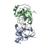
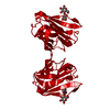
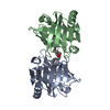
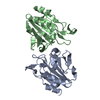
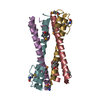
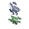
- PDB-1qxh: Crystal Structure of Escherichia coli Thiol Peroxidase in the Oxi... -
+ Open data

- Basic information
| Entry | Database: PDB / ID: 1qxh | ||||||
|---|---|---|---|---|---|---|---|
| Title | Crystal Structure of Escherichia coli Thiol Peroxidase in the Oxidized State | ||||||
 Components Components | Thiol peroxidase | ||||||
 Keywords Keywords |  OXIDOREDUCTASE / thiol peroxidase / scavengase P20 / antioxidant enzyme / peroxiredoxin OXIDOREDUCTASE / thiol peroxidase / scavengase P20 / antioxidant enzyme / peroxiredoxin | ||||||
| Function / homology |  Function and homology information Function and homology informationhydroperoxide reductase activity / thioredoxin-dependent peroxiredoxin / thioredoxin peroxidase activity / cellular response to oxidative stress / periplasmic space / cytosol / cytoplasmSimilarity search - Function | ||||||
| Biological species |  Escherichia coli (E. coli) Escherichia coli (E. coli) | ||||||
| Method | X-RAY DIFFRACTION / SYNCHROTRON / MIRAS / Resolution: 2.2 Å | ||||||
 Authors Authors | Choi, J. / Choi, S. / Choi, J. / Shin, W. | ||||||
 Citation Citation | Journal: J.Biol.Chem. / Year: 2003 Title: Crystal structure of Escherichia coli thiol peroxidase in the oxidized state: insights into intramolecular disulfide formation and substrate binding in atypical 2-Cys peroxiredoxins Authors: Choi, J. / Choi, S. / Choi, J. / Cha, M.-K. / Kim, I.-H. / Shin, W. #1: Journal: ACTA CRYSTALLOGR.,SECT.D / Year: 2003Title: Crystallization and preliminary X-ray analysis of Escherichia coli p20, a novel thiol peroxidase Authors: Choi, J. / Choi, S. / Choi, J. / Cha, M.-K. / Kim, I.-H. / Shin, W. | ||||||
| History |
|
- Structure visualization
| Structure viewer | Molecule: MolmilJmol/JSmol |
|---|
- Downloads & links
-Download
| PDBx/mmCIF format | 1qxh.cif.gz | 75.2 KB | Display | PDBx/mmCIF format |
|---|---|---|---|---|
| PDB format | pdb1qxh.ent.gz | 60.8 KB | Display | PDB format |
| PDBx/mmJSON format | 1qxh.json.gz | Tree view | PDBx/mmJSON format | |
| Others |  Other downloads Other downloads |
-Validation report
| Arichive directory | https://data.pdbj.org/pub/pdb/validation_reports/qx/1qxhftp://data.pdbj.org/pub/pdb/validation_reports/qx/1qxh | HTTPS FTP |
|---|
-Related structure data
| Similar structure data |
|---|


-Links
 PDBj
PDBj- Assembly
| Deposited unit | 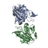
| ||||||||
|---|---|---|---|---|---|---|---|---|---|
| 1 |
| ||||||||
| Unit cell |
|
-Components
| #1: Protein | Mass: 17717.020 Da / Num. of mol.: 2 Source method: isolated from a genetically manipulated source Source: (gene. exp.) Escherichia coli (E. coli) / Plasmid: pT7-7 / Species (production host): Escherichia coli / Production host: Escherichia coli BL21 (bacteria) / Strain (production host): BL21References: UniProt: P0A862, Oxidoreductases; Acting on a peroxide as acceptor; Peroxidases#2: Water | ChemComp-HOH / | Water Mass: 18.015 Da / Num. of mol.: 335 / Source method: isolated from a natural source / Formula: H2O Mass: 18.015 Da / Num. of mol.: 335 / Source method: isolated from a natural source / Formula: H2O |
|---|
-Experimental details
-Experiment
| Experiment | Method: X-RAY DIFFRACTION / Number of used crystals: 1 |
|---|
- Sample preparation
| Crystal | Density Matthews: 1.85 Å3/Da / Density % sol: 33.01 % | ||||||||||||||||||||||||||||||
|---|---|---|---|---|---|---|---|---|---|---|---|---|---|---|---|---|---|---|---|---|---|---|---|---|---|---|---|---|---|---|---|
| Crystal grow | Temperature: 293 K / Method: vapor diffusion, hanging drop / pH: 7 Details: PEG 4000, iso-propyl alcohol, pH 7.0, VAPOR DIFFUSION, HANGING DROP, temperature 293K | ||||||||||||||||||||||||||||||
| Crystal grow | *PLUS pH: 7 | ||||||||||||||||||||||||||||||
| Components of the solutions | *PLUS
|
-Data collection
| Diffraction | Mean temperature: 100 K |
|---|---|
| Diffraction source | Source: SYNCHROTRON / Site: PAL/PLS  / Beamline: 6B / Wavelength: 1.12714 Å / Beamline: 6B / Wavelength: 1.12714 Å |
| Detector | Type: MACSCIENCE / Detector: IMAGE PLATE / Date: Jul 8, 2002 |
| Radiation | Monochromator: double crystal monochromator / Protocol: SINGLE WAVELENGTH / Monochromatic (M) / Laue (L): M / Scattering type: x-ray |
| Radiation wavelength | Wavelength: 1.12714 Å / Relative weight: 1 |
| Reflection | Resolution: 2.2→30 Å / Num. all: 15120 / Num. obs: 15120 / % possible obs: 99 % / Observed criterion σ(F): 0 / Observed criterion σ(I): 0 / Redundancy: 3.4 % / Biso Wilson estimate: 13.6 Å2 / Rmerge(I) obs: 0.063 / Rsym value: 0.063 / Net I/σ(I): 16.2 |
| Reflection shell | Resolution: 2.2→2.28 Å / Redundancy: 3.4 % / Mean I/σ(I) obs: 8.5 / Num. unique all: 1429 / Rsym value: 0.21 / % possible all: 99.4 |
| Reflection | *PLUS Lowest resolution: 30 Å / Num. measured all: 51963 |
| Reflection shell | *PLUS % possible obs: 99.4 % / Num. unique obs: 1429 / Num. measured obs: 4891 / Rmerge(I) obs: 0.21 |
- Processing
| Software |
| ||||||||||||||||||||||||||||||||||||||||||||||||||||||||||||
|---|---|---|---|---|---|---|---|---|---|---|---|---|---|---|---|---|---|---|---|---|---|---|---|---|---|---|---|---|---|---|---|---|---|---|---|---|---|---|---|---|---|---|---|---|---|---|---|---|---|---|---|---|---|---|---|---|---|---|---|---|---|
| Refinement | Method to determine structure: MIRAS / Resolution: 2.2→20.02 Å / Rfactor Rfree error: 0.009 / Data cutoff high absF: 930183.67 / Data cutoff low absF: 0 / Isotropic thermal model: RESTRAINED / Cross valid method: THROUGHOUT / σ(F): 0 / Stereochemistry target values: Engh & Huber
| ||||||||||||||||||||||||||||||||||||||||||||||||||||||||||||
| Solvent computation | Solvent model: FLAT MODEL / Bsol: 45.5007 Å2 / ksol: 0.332231 e/Å3 | ||||||||||||||||||||||||||||||||||||||||||||||||||||||||||||
| Displacement parameters | Biso mean: 30.2 Å2
| ||||||||||||||||||||||||||||||||||||||||||||||||||||||||||||
| Refine analyze |
| ||||||||||||||||||||||||||||||||||||||||||||||||||||||||||||
| Refinement step | Cycle: LAST / Resolution: 2.2→20.02 Å
| ||||||||||||||||||||||||||||||||||||||||||||||||||||||||||||
| Refine LS restraints |
| ||||||||||||||||||||||||||||||||||||||||||||||||||||||||||||
| LS refinement shell | Resolution: 2.2→2.34 Å / Rfactor Rfree error: 0.029 / Total num. of bins used: 6
| ||||||||||||||||||||||||||||||||||||||||||||||||||||||||||||
| Xplor file |
| ||||||||||||||||||||||||||||||||||||||||||||||||||||||||||||
| Refinement | *PLUS Rfactor obs: 0.188 / Rfactor Rfree: 0.246 / Rfactor Rwork: 0.188 | ||||||||||||||||||||||||||||||||||||||||||||||||||||||||||||
| Solvent computation | *PLUS | ||||||||||||||||||||||||||||||||||||||||||||||||||||||||||||
| Displacement parameters | *PLUS Biso mean: 30.3 Å2 | ||||||||||||||||||||||||||||||||||||||||||||||||||||||||||||
| Refine LS restraints | *PLUS
| ||||||||||||||||||||||||||||||||||||||||||||||||||||||||||||
| LS refinement shell | *PLUS Rfactor Rfree: 0.28 |
