 Movie
Movie Controller


+ Open data

- Basic information
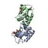
| Entry | Database: PDB / ID: 1q98 | ||||||
|---|---|---|---|---|---|---|---|
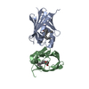
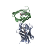
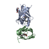
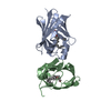
| Title | Structure of a Thiol Peroxidase from Haemophilus influenzae Rd | ||||||
 Components Components | Thiol Peroxidase | ||||||
 Keywords Keywords |  OXIDOREDUCTASE / structural genomics / tpx / peroxidase / NYSGXRC / T1429 / PSI / Protein Structure Initiative / New York SGX Research Center for Structural Genomics OXIDOREDUCTASE / structural genomics / tpx / peroxidase / NYSGXRC / T1429 / PSI / Protein Structure Initiative / New York SGX Research Center for Structural Genomics | ||||||
| Function / homology |  Function and homology information Function and homology information | ||||||
| Biological species |  Haemophilus influenzae (bacteria) Haemophilus influenzae (bacteria) | ||||||
| Method | X-RAY DIFFRACTION / SYNCHROTRON / SIRAS ON HG / Resolution: 1.9 Å | ||||||
 Authors Authors | Kniewel, R. / Buglino, J. / Solorzano, V. / Wu, J. / Lima, C.D. / Burley, S.K. / New York SGX Research Center for Structural Genomics (NYSGXRC) | ||||||
 Citation Citation | Journal: To be Published Title: Structure of a Thiol Peroxidase from Haemophilus influenzae Rd Authors: Kniewel, R. / Buglino, J. / Solorzano, V. / Wu, J. / Lima, C.D. | ||||||
| History |
|
- Structure visualization
| Structure viewer | Molecule: MolmilJmol/JSmol |
|---|
- Downloads & links
-Download
| PDBx/mmCIF format | 1q98.cif.gz | 75.1 KB | Display | PDBx/mmCIF format |
|---|---|---|---|---|
| PDB format | pdb1q98.ent.gz | 60 KB | Display | PDB format |
| PDBx/mmJSON format | 1q98.json.gz | Tree view | PDBx/mmJSON format | |
| Others |  Other downloads Other downloads |
-Validation report
| Arichive directory | https://data.pdbj.org/pub/pdb/validation_reports/q9/1q98ftp://data.pdbj.org/pub/pdb/validation_reports/q9/1q98 | HTTPS FTP |
|---|
-Related structure data
| Similar structure data | |
|---|---|
| Other databases |

-Links
 PDBj
PDBj- Assembly
| Deposited unit | 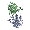
| ||||||||||
|---|---|---|---|---|---|---|---|---|---|---|---|
| 1 |
| ||||||||||
| Unit cell |
|
-Components
| #1: Protein | Mass: 17640.990 Da / Num. of mol.: 2 Source method: isolated from a genetically manipulated source Source: (gene. exp.) Haemophilus influenzae (bacteria) / Gene: tpx, HI0751 / Plasmid: pET28b / Species (production host): Escherichia coli / Production host: Escherichia coli BL21(DE3) (bacteria) / Strain (production host): BL21 DE3References: UniProt: Q57549, Oxidoreductases; Acting on a peroxide as acceptor; Peroxidases#2: Water | ChemComp-HOH / | Water Mass: 18.015 Da / Num. of mol.: 275 / Source method: isolated from a natural source / Formula: H2O Mass: 18.015 Da / Num. of mol.: 275 / Source method: isolated from a natural source / Formula: H2O |
|---|
-Experimental details
-Experiment
| Experiment | Method: X-RAY DIFFRACTION / Number of used crystals: 2 |
|---|
- Sample preparation
| Crystal | Density Matthews: 2.07 Å3/Da / Density % sol: 40.1 % |
|---|---|
| Crystal grow | Temperature: 291 K / Method: vapor diffusion, hanging drop / pH: 4.5 Details: 19-24% PEGv 4000, 0.1M Ammonium Acetate, 0.1M Sodium Acetate pH 4.5, 5%Ethylene Glycol, VAPOR DIFFUSION, HANGING DROP, temperature 291K |
-Data collection
| Diffraction | Mean temperature: 100 K |
|---|---|
| Diffraction source | Source: SYNCHROTRON / Site: APS  / Beamline: 31-ID / Wavelength: 0.9798 Å / Beamline: 31-ID / Wavelength: 0.9798 Å |
| Detector | Type: MARRESEARCH / Detector: CCD / Date: Jun 27, 2003 |
| Radiation | Monochromator: Si 111 CHANNEL / Protocol: SINGLE WAVELENGTH / Monochromatic (M) / Laue (L): M / Scattering type: x-ray |
| Radiation wavelength | Wavelength: 0.9798 Å / Relative weight: 1 |
| Reflection | Resolution: 1.9→20 Å / Num. all: 24969 / Num. obs: 24220 / % possible obs: 97 % / Observed criterion σ(I): -3 / Redundancy: 4 % / Biso Wilson estimate: 26.6 Å2 / Rmerge(I) obs: 0.039 / Net I/σ(I): 7.9 |
| Reflection shell | Resolution: 1.9→1.97 Å / Redundancy: 4 % / Rmerge(I) obs: 0.162 / Num. unique all: 2399 / % possible all: 96.4 |
- Processing
| Software |
| ||||||||||||||||||||||||||||||||||||
|---|---|---|---|---|---|---|---|---|---|---|---|---|---|---|---|---|---|---|---|---|---|---|---|---|---|---|---|---|---|---|---|---|---|---|---|---|---|
| Refinement | Method to determine structure: SIRAS ON HG / Resolution: 1.9→18.91 Å / Rfactor Rfree error: 0.008 / Isotropic thermal model: RESTRAINED / Cross valid method: THROUGHOUT / σ(F): 0 / Stereochemistry target values: Engh & Huber / Details: BULK SOLVENT MODEL USED
| ||||||||||||||||||||||||||||||||||||
| Solvent computation | Solvent model: FLAT MODEL / Bsol: 62.7964 Å2 / ksol: 0.372079 e/Å3 | ||||||||||||||||||||||||||||||||||||
| Displacement parameters | Biso mean: 37.6 Å2
| ||||||||||||||||||||||||||||||||||||
| Refine analyze | Luzzati coordinate error free: 0.3 Å / Luzzati sigma a free: 0.2 Å | ||||||||||||||||||||||||||||||||||||
| Refinement step | Cycle: LAST / Resolution: 1.9→18.91 Å
| ||||||||||||||||||||||||||||||||||||
| Refine LS restraints |
| ||||||||||||||||||||||||||||||||||||
| LS refinement shell | Resolution: 1.9→2.02 Å / Rfactor Rfree error: 0.022 / Total num. of bins used: 6
| ||||||||||||||||||||||||||||||||||||
| Xplor file |
|
