 Movie
Movie Controller


+ Open data

- Basic information
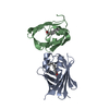
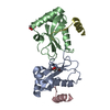
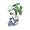
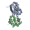
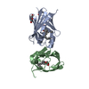
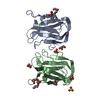
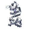
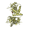
| Entry | Database: PDB / ID: 1k1f | ||||||
|---|---|---|---|---|---|---|---|
| Title | Structure of the Bcr-Abl Oncoprotein Oligomerization domain | ||||||
 Components Components | BREAKPOINT CLUSTER REGION PROTEIN | ||||||
 Keywords Keywords |  TRANSFERASE / Oligomerization / coiled coil / Bcr-Abl kinase TRANSFERASE / Oligomerization / coiled coil / Bcr-Abl kinase | ||||||
| Function / homology |  Function and homology information Function and homology informationnegative regulation of respiratory burst / negative regulation of cellular extravasation / negative regulation of macrophage migration / : / negative regulation of blood vessel remodeling / negative regulation of neutrophil degranulation / macrophage migration / neutrophil degranulation / intracellular protein transmembrane transport / renal system process ...negative regulation of respiratory burst / negative regulation of cellular extravasation / negative regulation of macrophage migration / : / negative regulation of blood vessel remodeling / negative regulation of neutrophil degranulation / macrophage migration / neutrophil degranulation / intracellular protein transmembrane transport / renal system process / regulation of vascular permeability / regulation of Rho protein signal transduction / focal adhesion assembly / definitive hemopoiesis / Signaling by cytosolic FGFR1 fusion mutants / activation of GTPase activity / regulation of small GTPase mediated signal transduction / inner ear morphogenesis / small GTPase-mediated signal transduction / RHOB GTPase cycle / RHOC GTPase cycle / CDC42 GTPase cycle / neuromuscular process controlling balance / homeostasis of number of cells / RHOA GTPase cycle / negative regulation of reactive oxygen species metabolic process / RAC2 GTPase cycle / RAC3 GTPase cycle / phagocytosis / positive regulation of phagocytosis / keratinocyte differentiation / RAC1 GTPase cycle / Signaling by FGFR1 in disease / GTPase activator activity / guanyl-nucleotide exchange factor activity / Schaffer collateral - CA1 synapse / brain development / modulation of chemical synaptic transmission / negative regulation of inflammatory response / actin cytoskeleton organization / protein tyrosine kinase activity / cellular response to lipopolysaccharide / dendritic spine / postsynaptic density / non-specific serine/threonine protein kinase / regulation of cell cycle / axon / protein phosphorylation / protein serine kinase activity / protein serine/threonine kinase activity / glutamatergic synapse / signal transduction / protein-containing complex / extracellular exosome / ATP binding / membrane / plasma membrane / cytosolSimilarity search - Function | ||||||
| Biological species |  Homo sapiens (human) Homo sapiens (human) | ||||||
| Method | X-RAY DIFFRACTION / SYNCHROTRON / MAD / Resolution: 2.2 Å | ||||||
 Authors Authors | Zhao, X. / Ghaffari, S. / Lodish, H. / Malashkevich, V.N. / Kim, P.S. | ||||||
 Citation Citation | Journal: Nat.Struct.Biol. / Year: 2002 Title: Structure of the Bcr-Abl oncoprotein oligomerization domain. Authors: Zhao, X. / Ghaffari, S. / Lodish, H. / Malashkevich, V.N. / Kim, P.S. | ||||||
| History |
|
- Structure visualization
| Structure viewer | Molecule: MolmilJmol/JSmol |
|---|
- Downloads & links
-Download
| PDBx/mmCIF format | 1k1f.cif.gz | 122 KB | Display | PDBx/mmCIF format |
|---|---|---|---|---|
| PDB format | pdb1k1f.ent.gz | 104.1 KB | Display | PDB format |
| PDBx/mmJSON format | 1k1f.json.gz | Tree view | PDBx/mmJSON format | |
| Others |  Other downloads Other downloads |
-Validation report
| Arichive directory | https://data.pdbj.org/pub/pdb/validation_reports/k1/1k1fftp://data.pdbj.org/pub/pdb/validation_reports/k1/1k1f | HTTPS FTP |
|---|
-Related structure data
| Similar structure data |
|---|


-Links
 PDBj
PDBj


- Assembly
| Deposited unit | 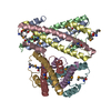
| ||||||||
|---|---|---|---|---|---|---|---|---|---|
| 1 | 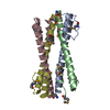
| ||||||||
| 2 | 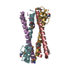
| ||||||||
| Unit cell |
|
-Components
| #1: Protein | Mass: 8709.438 Da / Num. of mol.: 8 / Fragment: bcr1-72 / Mutation: C38A Source method: isolated from a genetically manipulated source Source: (gene. exp.) Homo sapiens (human) / Production host:  Escherichia coli (E. coli) Escherichia coli (E. coli)References: UniProt: P11274, Transferases; Transferring phosphorus-containing groups; Phosphotransferases with an alcohol group as acceptor#2: Water | ChemComp-HOH / | Water Mass: 18.015 Da / Num. of mol.: 420 / Source method: isolated from a natural source / Formula: H2O Mass: 18.015 Da / Num. of mol.: 420 / Source method: isolated from a natural source / Formula: H2O |
|---|
-Experimental details
-Experiment
| Experiment | Method: X-RAY DIFFRACTION / Number of used crystals: 2 |
|---|
- Sample preparation
| Crystal | Density Matthews: 1.89 Å3/Da / Density % sol: 34.84 % | ||||||||||||||||||||||||||||||
|---|---|---|---|---|---|---|---|---|---|---|---|---|---|---|---|---|---|---|---|---|---|---|---|---|---|---|---|---|---|---|---|
| Crystal grow | Temperature: 293 K / Method: vapor diffusion, hanging drop / pH: 5 Details: ammonium sulfate, pH 5.0, VAPOR DIFFUSION, HANGING DROP, temperature 293K | ||||||||||||||||||||||||||||||
| Crystal grow | *PLUS pH: 6.5 / Method: vapor diffusion | ||||||||||||||||||||||||||||||
| Components of the solutions | *PLUS
|
-Data collection
| Diffraction |
| ||||||||||||
|---|---|---|---|---|---|---|---|---|---|---|---|---|---|
| Diffraction source | Source: SYNCHROTRON / Site: NSLS  / Beamline: X4A / Wavelength: 0.9686,0.9789,0.9793 / Beamline: X4A / Wavelength: 0.9686,0.9789,0.9793 | ||||||||||||
| Detector | Type: ADSC QUANTUM 4 / Detector: CCD / Date: Mar 7, 2001 | ||||||||||||
| Radiation | Monochromator: Graphite / Protocol: MAD / Monochromatic (M) / Laue (L): M / Scattering type: x-ray | ||||||||||||
| Radiation wavelength |
| ||||||||||||
| Reflection | Resolution: 2.2→20 Å / Num. all: 51720 / Num. obs: 51251 / % possible obs: 99 % / Observed criterion σ(F): 2 / Observed criterion σ(I): 2 / Biso Wilson estimate: 27.1 Å2 | ||||||||||||
| Reflection | *PLUS Lowest resolution: 20 Å / % possible obs: 99 % |
- Processing
| Software |
| |||||||||||||||||||||||||
|---|---|---|---|---|---|---|---|---|---|---|---|---|---|---|---|---|---|---|---|---|---|---|---|---|---|---|
| Refinement | Method to determine structure: MAD / Resolution: 2.2→10 Å / Data cutoff high absF: 1412713.36 / Data cutoff low absF: 0 / Isotropic thermal model: RESTRAINED / Cross valid method: THROUGHOUT / σ(F): 0 / Stereochemistry target values: Engh & Huber
| |||||||||||||||||||||||||
| Solvent computation | Solvent model: FLAT MODEL / Bsol: 80.6333 Å2 / ksol: 0.477025 e/Å3 | |||||||||||||||||||||||||
| Displacement parameters | Biso mean: 40.5 Å2
| |||||||||||||||||||||||||
| Refine analyze | Luzzati coordinate error obs: 0.36 Å / Luzzati d res low obs: 5 Å / Luzzati sigma a obs: 0.28 Å | |||||||||||||||||||||||||
| Refinement step | Cycle: LAST / Resolution: 2.2→10 Å
| |||||||||||||||||||||||||
| Refine LS restraints |
| |||||||||||||||||||||||||
| LS refinement shell | Resolution: 2.4→2.55 Å / Total num. of bins used: 6
| |||||||||||||||||||||||||
| Software | *PLUS Name: CNS / Version: 1 / Classification: refinement | |||||||||||||||||||||||||
| Refinement | *PLUS Lowest resolution: 10 Å / σ(F): 0 | |||||||||||||||||||||||||
| Solvent computation | *PLUS | |||||||||||||||||||||||||
| Displacement parameters | *PLUS Biso mean: 40.5 Å2 | |||||||||||||||||||||||||
| Refine LS restraints | *PLUS
| |||||||||||||||||||||||||
| LS refinement shell | *PLUS Highest resolution: 2.15 Å / Lowest resolution: 2.2 Å / Rfactor Rfree: 0.387 / Rfactor Rwork: 0.308 / Rfactor obs: 0.308 |
