 Movie
Movie Controller


+ Open data

- Basic information
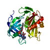
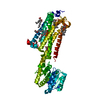
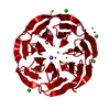
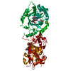
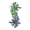
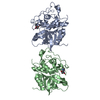
| Entry | Database: PDB / ID: 1qx4 | ||||||
|---|---|---|---|---|---|---|---|
| Title | Structrue of S127P mutant of cytochrome b5 reductase | ||||||
 Components Components | NADH-cytochrome b5 reductase | ||||||
 Keywords Keywords |  OXIDOREDUCTASE / methemoglobinemia / flavin flexibility OXIDOREDUCTASE / methemoglobinemia / flavin flexibility | ||||||
| Function / homology |  Function and homology information Function and homology informationPhase I - Functionalization of compounds / nitric-oxide synthase complex / Vitamin C (ascorbate) metabolism / cytochrome-b5 reductase / cytochrome-b5 reductase activity, acting on NAD(P)H / nitrite reductase (NO-forming) activity / Neutrophil degranulation / AMP binding / cholesterol biosynthetic process / nitric oxide biosynthetic process ...Phase I - Functionalization of compounds / nitric-oxide synthase complex / Vitamin C (ascorbate) metabolism / cytochrome-b5 reductase / cytochrome-b5 reductase activity, acting on NAD(P)H / nitrite reductase (NO-forming) activity / Neutrophil degranulation / AMP binding / cholesterol biosynthetic process / nitric oxide biosynthetic process / FAD binding / lipid droplet / mitochondrial membrane / ADP binding / NAD binding / flavin adenine dinucleotide binding / mitochondrial outer membrane / endoplasmic reticulum membrane / mitochondrionSimilarity search - Function | ||||||
| Biological species |  Rattus norvegicus (Norway rat) Rattus norvegicus (Norway rat) | ||||||
| Method | X-RAY DIFFRACTION / SYNCHROTRON / MOLECULAR REPLACEMENT / Resolution: 1.8 Å | ||||||
 Authors Authors | Bewley, M.C. / Davis, C.A. / Marohnic, C.C. / Taormina, D. / Barber, M.J. | ||||||
 Citation Citation | Journal: Biochemistry / Year: 2003 Title: The structure of the S127P mutant of cytochrome b5 reductase that causes methemoglobinemia shows the AMP moiety of the flavin occupying the substrate binding site Authors: Bewley, M.C. / Davis, C.A. / Marohnic, C.C. / Taormina, D. / Barber, M.J. | ||||||
| History |
|
- Structure visualization
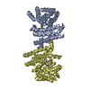
| Structure viewer | Molecule: MolmilJmol/JSmol |
|---|
- Downloads & links
-Download
| PDBx/mmCIF format | 1qx4.cif.gz | 123.9 KB | Display | PDBx/mmCIF format |
|---|---|---|---|---|
| PDB format | pdb1qx4.ent.gz | 94.9 KB | Display | PDB format |
| PDBx/mmJSON format | 1qx4.json.gz | Tree view | PDBx/mmJSON format | |
| Others |  Other downloads Other downloads |
-Validation report
| Arichive directory | https://data.pdbj.org/pub/pdb/validation_reports/qx/1qx4ftp://data.pdbj.org/pub/pdb/validation_reports/qx/1qx4 | HTTPS FTP |
|---|
-Related structure data
| Related structure data |  1ib0S S: Starting model for refinement |
|---|---|
| Similar structure data |


-Links
 PDBj
PDBj


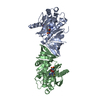
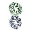
- Assembly
| Deposited unit | 
| ||||||||
|---|---|---|---|---|---|---|---|---|---|
| 1 |
| ||||||||
| Unit cell |
|
-Components
| #1: Protein | Mass: 31325.217 Da / Num. of mol.: 2 / Mutation: S127P Source method: isolated from a genetically manipulated source Source: (gene. exp.) Rattus norvegicus (Norway rat) / Gene: DIA1 / Production host:  Escherichia coli (E. coli) / Strain (production host): BL21-Codon-Plus (DE3)RIL / References: UniProt: P20070, cytochrome-b5 reductase Escherichia coli (E. coli) / Strain (production host): BL21-Codon-Plus (DE3)RIL / References: UniProt: P20070, cytochrome-b5 reductase#2: Chemical | Flavin adenine dinucleotide  Mass: 785.550 Da / Num. of mol.: 2 / Source method: obtained synthetically / Formula: C27H33N9O15P2 / Comment: FAD*YM Mass: 785.550 Da / Num. of mol.: 2 / Source method: obtained synthetically / Formula: C27H33N9O15P2 / Comment: FAD*YM#3: Water | ChemComp-HOH / | Water Mass: 18.015 Da / Num. of mol.: 256 / Source method: isolated from a natural source / Formula: H2O Mass: 18.015 Da / Num. of mol.: 256 / Source method: isolated from a natural source / Formula: H2O |
|---|
-Experimental details
-Experiment
| Experiment | Method: X-RAY DIFFRACTION |
|---|
- Sample preparation
| Crystal | Density Matthews: 2.18 Å3/Da / Density % sol: 43.52 % |
|---|---|
| Crystal grow | Temperature: 298 K / Method: vapor diffusion, sitting drop / pH: 8.5 Details: glycerol, ammonium sulfate, tris buffer, pH 8.5, VAPOR DIFFUSION, SITTING DROP, temperature 298.0K |
-Data collection
| Diffraction | Mean temperature: 99 K |
|---|---|
| Diffraction source | Source: SYNCHROTRON / Site: NSLS  / Beamline: X12B / Wavelength: 0.98 Å / Beamline: X12B / Wavelength: 0.98 Å |
| Detector | Type: ADSC QUANTUM 4 / Detector: CCD / Date: May 15, 2001 |
| Radiation | Monochromator: graphite / Protocol: SINGLE WAVELENGTH / Monochromatic (M) / Laue (L): M / Scattering type: x-ray |
| Radiation wavelength | Wavelength: 0.98 Å / Relative weight: 1 |
| Reflection | Resolution: 1.8→30 Å / Num. all: 48325 / Num. obs: 48325 / % possible obs: 96 % / Rmerge(I) obs: 0.064 |
- Processing
| Software |
| ||||||||||||||||||||||||||||
|---|---|---|---|---|---|---|---|---|---|---|---|---|---|---|---|---|---|---|---|---|---|---|---|---|---|---|---|---|---|
| Refinement | Method to determine structure: MOLECULAR REPLACEMENT Starting model: PDB ENTRY 1IB0 without FAD molecule Resolution: 1.8→30 Å / Cross valid method: THROUGHOUT / Stereochemistry target values: Engh & Huber
| ||||||||||||||||||||||||||||
| Solvent computation | Bsol: 24.0223 Å2 / ksol: 0.438183 e/Å3 | ||||||||||||||||||||||||||||
| Displacement parameters |
| ||||||||||||||||||||||||||||
| Refinement step | Cycle: LAST / Resolution: 1.8→30 Å
| ||||||||||||||||||||||||||||
| Refine LS restraints |
|
