 Movie
Movie Controller

[English] 日本語
 Yorodumi
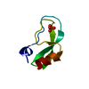
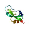
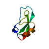
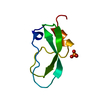
- PDB-1qlq: Bovine Pancreatic Trypsin Inhibitor (BPTI) Mutant with Altered Bi... -
+ Open data

- Basic information
| Entry | Database: PDB / ID: 1qlq | ||||||
|---|---|---|---|---|---|---|---|
| Title | Bovine Pancreatic Trypsin Inhibitor (BPTI) Mutant with Altered Binding Loop Sequence | ||||||
 Components Components | PANCREATIC TRYPSIN INHIBITOR | ||||||
 Keywords Keywords |  SERINE PROTEASE INHIBITOR SERINE PROTEASE INHIBITOR | ||||||
| Function / homology |  Function and homology information Function and homology informationtrypsinogen activation / negative regulation of serine-type endopeptidase activity / sulfate binding / potassium channel inhibitor activity / negative regulation of platelet aggregation / zymogen binding / molecular function inhibitor activity / negative regulation of thrombin-activated receptor signaling pathway / serine protease inhibitor complex / serine-type endopeptidase inhibitor activity ...trypsinogen activation / negative regulation of serine-type endopeptidase activity / sulfate binding / potassium channel inhibitor activity / negative regulation of platelet aggregation / zymogen binding / molecular function inhibitor activity / negative regulation of thrombin-activated receptor signaling pathway / serine protease inhibitor complex / serine-type endopeptidase inhibitor activity / protease binding / calcium ion binding / extracellular spaceSimilarity search - Function | ||||||
| Biological species |  BOS TAURUS (cattle) BOS TAURUS (cattle) | ||||||
| Method | X-RAY DIFFRACTION / MOLECULAR REPLACEMENT / Resolution: 1.42 Å | ||||||
 Authors Authors | Czapinska, H. / Krzywda, S. / Sheldrick, G.M. / Otlewski, J. / Jaskolski, M. | ||||||
 Citation Citation | Journal: J.Mol.Biol. / Year: 1999 Title: High Resolution Structure of Bovine Pancreatic Trypsin Inhibitor with Altered Binding Loop Sequence Authors: Czapinska, H. / Otlewski, J. / Krzywda, S. / Sheldrick, G.M. / Jaskolski, M. #1: Journal: Acta Crystallogr.,Sect.D / Year: 1996Title: Structure of Bovine Pancreatic Trypsin Inhibitor at 125 K: Definition of Carboxyl-Terminal Residues Gly57 and Ala58 Authors: Parkin, S. / Rupp, B. / Hope, H. #2: Journal: J.Mol.Biol. / Year: 1992Title: Determination of a High Quality Nuclear Magnetic Resonance Solution Structure of the Bovine Pancreatic Trypsin Inhibitor and Comparison with Three Crystal Structures Authors: Berndt, K. / Guentert, P. / Orbons, L.P. / Wuethrich, K. #3: Journal: J.Mol.Biol. / Year: 1984Title: Structure of Bovine Pancreatic Trypsin Inhibitor . Results of Joint Neutron and X-Ray Refinement of Crystal Form II Authors: Wlodawer, A. / Walter, J. / Huber, R. / Sjolin, L. #4: Journal: Acta Crystallogr.,Sect.B / Year: 1975Title: Crystallographic Refinement of the Structure of Bovine Pancreatic Trypsin Inhibitor at 1.5 A Resolution Authors: Deisenhofer, J. / Steigemann, W. | ||||||
| History |
|
- Structure visualization
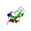
| Structure viewer | Molecule: MolmilJmol/JSmol |
|---|
- Downloads & links
-Download
| PDBx/mmCIF format | 1qlq.cif.gz | 44.2 KB | Display | PDBx/mmCIF format |
|---|---|---|---|---|
| PDB format | pdb1qlq.ent.gz | 31.1 KB | Display | PDB format |
| PDBx/mmJSON format | 1qlq.json.gz | Tree view | PDBx/mmJSON format | |
| Others |  Other downloads Other downloads |
-Validation report
| Arichive directory | https://data.pdbj.org/pub/pdb/validation_reports/ql/1qlqftp://data.pdbj.org/pub/pdb/validation_reports/ql/1qlq | HTTPS FTP |
|---|
-Related structure data
| Related structure data |  1bpiS S: Starting model for refinement |
|---|---|
| Similar structure data |


-Links
 PDBj
PDBj


- Assembly
| Deposited unit | 
| |||||||||
|---|---|---|---|---|---|---|---|---|---|---|
| 1 |
| |||||||||
| Unit cell |
| |||||||||
| Components on special symmetry positions |
| |||||||||
| Details | THE ASYMMETRIC UNIT CONTAINS THE MONOMERIC MOLECULE.AN EXTRA WATER MOLECULE IS BURIED IN AN INTERNAL CLEFT.IN THE CRYSTAL THE ARG17-ILE18- ILE19 SEGMENTS OF THE PROTEINMOLECULES RELATED BY THE (Y, X, -Z) TWO-FOLD ROTATION FORMAN INTERMOLECULAR ANTIPARALLEL B-SHEET.A DIFFERENT INTERMOLECULAR ANTIPARALLEL B-SHEET IS FOUNDIN THE CRYSTAL FOR THE BPTI 8PTI WHERE IN SPACE GROUPP 42 21 2, STRAND 31 TO 35 IS INVOLVED IN A CRYSTAL PACKINGDIMER. FOR THE BPTI STRUCTURES 6PTI AND 1NAG THE LOOP39 TO 42 IS INVOLVED IN A DIFFERENT TIGHT CRYSTALPACKING, SPACE GROUP P 21 21 2, WHILE A DECAMER IS OBSERVEDFOR THE BPTI IN ENTRIES 1B0C AND 1BZ5. |
-Components
| #1: Protein | Mass: 6481.481 Da / Num. of mol.: 1 / Mutation: YES Source method: isolated from a genetically manipulated source Source: (gene. exp.) BOS TAURUS (cattle) / Organ: PANCREAS / Production host:  ESCHERICHIA COLI (E. coli) / Strain (production host): BL21(DE3) / Variant (production host): T7 / References: UniProt: P00974 ESCHERICHIA COLI (E. coli) / Strain (production host): BL21(DE3) / Variant (production host): T7 / References: UniProt: P00974 | ||
|---|---|---|---|
| #2: Chemical | ChemComp-SO4 / Sulfate  Mass: 96.063 Da / Num. of mol.: 4 / Source method: obtained synthetically / Formula: SO4 Mass: 96.063 Da / Num. of mol.: 4 / Source method: obtained synthetically / Formula: SO4#3: Water | ChemComp-HOH / | Water Mass: 18.015 Da / Num. of mol.: 98 / Source method: isolated from a natural source / Formula: H2O Mass: 18.015 Da / Num. of mol.: 98 / Source method: isolated from a natural source / Formula: H2O |
-Experimental details
-Experiment
| Experiment | Method: X-RAY DIFFRACTION / Number of used crystals: 1 |
|---|
- Sample preparation
| Crystal | Density Matthews: 2.36 Å3/Da / Density % sol: 48 % | |||||||||||||||||||||||||
|---|---|---|---|---|---|---|---|---|---|---|---|---|---|---|---|---|---|---|---|---|---|---|---|---|---|---|
| Crystal grow | Temperature: 292 K / Method: vapor diffusion, hanging drop / pH: 7.5 Details: A PROTEIN SAMPLE, LYOPHILIZED AFTER HPLC PURIFICATION FROM TFA/ACETONITRILE MIXTURE, WAS DISSOLVED IN WATER TO A CONCENTRATION OF 9 MG/ML. 2 UL DROPS OF THE PROTEIN SOLUTION WERE MIXED WITH ...Details: A PROTEIN SAMPLE, LYOPHILIZED AFTER HPLC PURIFICATION FROM TFA/ACETONITRILE MIXTURE, WAS DISSOLVED IN WATER TO A CONCENTRATION OF 9 MG/ML. 2 UL DROPS OF THE PROTEIN SOLUTION WERE MIXED WITH 2 UL OF RESERVOIR SOLUTION CONTAINING 2% PEG 400, 2 M AMMONIUM SULFATE AND 0.1 M NA HEPES, PH 7.5. THE HANGING DROPLETS WERE EQUILIBRATED AT 19 DEG C THROUGH THE GAS PHASE WITH THE RESERVOIR. PRISMATIC CRYSTALS MEASURING UP TO 0.4 MM GREW WITHIN 12 HOURS. | |||||||||||||||||||||||||
| Crystal grow | *PLUS Temperature: 19 ℃ / Method: vapor diffusion, hanging drop / pH: 7 | |||||||||||||||||||||||||
| Components of the solutions | *PLUS
|
-Data collection
| Diffraction | Mean temperature: 290 K |
|---|---|
| Diffraction source | Source: ROTATING ANODE / Type: SIEMENS SRA2 / Wavelength: 1.5418 |
| Detector | Type: MARRESEARCH / Detector: IMAGE PLATE / Date: Jan 15, 1999 |
| Radiation | Monochromator: GRAPHITE(002) / Protocol: SINGLE WAVELENGTH / Monochromatic (M) / Laue (L): M / Scattering type: x-ray |
| Radiation wavelength | Wavelength: 1.5418 Å / Relative weight: 1 |
| Reflection | Resolution: 1.42→20 Å / Num. obs: 11934 / % possible obs: 99.6 % / Redundancy: 19.3 % / Rmerge(I) obs: 0.059 / Net I/σ(I): 50.2 |
| Reflection shell | Resolution: 1.42→1.47 Å / Redundancy: 6 % / Rmerge(I) obs: 0.326 / Mean I/σ(I) obs: 5.144 / % possible all: 96 |
| Reflection shell | *PLUS % possible obs: 96 % / Mean I/σ(I) obs: 5.1 |
- Processing
| Software |
| |||||||||||||||||||||||||||||||||
|---|---|---|---|---|---|---|---|---|---|---|---|---|---|---|---|---|---|---|---|---|---|---|---|---|---|---|---|---|---|---|---|---|---|---|
| Refinement | Method to determine structure: MOLECULAR REPLACEMENT Starting model: 1BPI Resolution: 1.42→10 Å / Num. parameters: 5297 / Num. restraintsaints: 5730 / Cross valid method: FREE R-VALUE / σ(F): 0 / Stereochemistry target values: ENGH AND HUBER Details: ANISOTROPIC REFINEMENT. THE COMPLETE C-TERMINUS IS VISIBLE. IT FORMS A SALT-BRIDGE WITH THE N- TERMINUS. THE CYS14-CYS38 DISULFIDE BRIDGE IS OBSERVED IN TWO DISTINCT CHIRALITIES (63 % RIGHT- ...Details: ANISOTROPIC REFINEMENT. THE COMPLETE C-TERMINUS IS VISIBLE. IT FORMS A SALT-BRIDGE WITH THE N- TERMINUS. THE CYS14-CYS38 DISULFIDE BRIDGE IS OBSERVED IN TWO DISTINCT CHIRALITIES (63 % RIGHT-HANDED, 37 % LEFT-HANDED). ONE TWO-FOLD SYMMETRIC AND THREE GENERAL-POSITION SULFATE ANIONS ARE PRESENT PER ONE PROTEIN MOLECULE.
| |||||||||||||||||||||||||||||||||
| Solvent computation | Solvent model: MOEWS & KRETSINGER | |||||||||||||||||||||||||||||||||
| Refine analyze | Num. disordered residues: 4 / Occupancy sum hydrogen: 429 / Occupancy sum non hydrogen: 544.5 | |||||||||||||||||||||||||||||||||
| Refinement step | Cycle: LAST / Resolution: 1.42→10 Å
| |||||||||||||||||||||||||||||||||
| Refine LS restraints |
| |||||||||||||||||||||||||||||||||
| Software | *PLUS Name: SHELXL-97 / Classification: refinement | |||||||||||||||||||||||||||||||||
| Refinement | *PLUS Rfactor all: 0.1087 / Rfactor obs: 0.1048 | |||||||||||||||||||||||||||||||||
| Solvent computation | *PLUS | |||||||||||||||||||||||||||||||||
| Displacement parameters | *PLUS | |||||||||||||||||||||||||||||||||
| Refine LS restraints | *PLUS
|
