 Movie
Movie Controller

[English] 日本語
 Yorodumi
- PDB-8pti: CRYSTAL STRUCTURE OF A Y35G MUTANT OF BOVINE PANCREATIC TRYPSIN I... -
+ Open data

- Basic information
| Entry | Database: PDB / ID: 8pti | ||||||
|---|---|---|---|---|---|---|---|
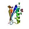
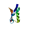
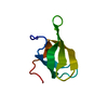
| Title | CRYSTAL STRUCTURE OF A Y35G MUTANT OF BOVINE PANCREATIC TRYPSIN INHIBITOR | ||||||
 Components Components | BOVINE PANCREATIC TRYPSIN INHIBITOR Aprotinin Aprotinin | ||||||
 Keywords Keywords | PROTEINASE INHIBITOR (TRYPSIN) | ||||||
| Function / homology |  Function and homology information Function and homology informationtrypsinogen activation / negative regulation of serine-type endopeptidase activity / sulfate binding / potassium channel inhibitor activity / negative regulation of platelet aggregation / zymogen binding / molecular function inhibitor activity / negative regulation of thrombin-activated receptor signaling pathway / serine protease inhibitor complex / serine-type endopeptidase inhibitor activity ...trypsinogen activation / negative regulation of serine-type endopeptidase activity / sulfate binding / potassium channel inhibitor activity / negative regulation of platelet aggregation / zymogen binding / molecular function inhibitor activity / negative regulation of thrombin-activated receptor signaling pathway / serine protease inhibitor complex / serine-type endopeptidase inhibitor activity / protease binding / calcium ion binding / extracellular spaceSimilarity search - Function | ||||||
| Biological species |  Bos taurus (cattle) Bos taurus (cattle) | ||||||
| Method | X-RAY DIFFRACTION / Resolution: 1.8 Å | ||||||
 Authors Authors | Housset, D. / Kim, K.-S. / Fuchs, J. / Woodward, C. / Wlodawer, A. | ||||||
 Citation Citation | Journal: J.Mol.Biol. / Year: 1991 Title: Crystal structure of a Y35G mutant of bovine pancreatic trypsin inhibitor. Authors: Housset, D. / Kim, K.S. / Fuchs, J. / Woodward, C. / Wlodawer, A. #1: Journal: Protein Eng. / Year: 1990Title: Structural Effects Induced by Removal of a Disulfide-Bridge. The X-Ray Structure of the C30A(Slash)C51A Mutant of Basic Pancreatic Trypsin Inhibitor at 1.6 Angstroms Authors: Eigenbrot, C. / Randal, M. / Kossiakoff, A.A. #2: Journal: Biochemistry / Year: 1990Title: X-Ray Crystal Structure of the Protease Inhibitor Domain of Alzheimer'S Amyloid Beta-Protein Precursor Authors: Hynes, T.R. / Randal, M. / Kennedy, L.A. / Eigenbrot, C. / Kossiakoff, A.A. #3: Journal: J.Mol.Biol. / Year: 1987Title: Structure of Form III Crystals of Bovine Pancreatic Trypsin Inhibitor. Authors: Wlodawer, A. / Nachman, J. / Gilliland, G.L. / Gallagher, W. / Woodward, C. #4: Journal: J.Mol.Biol. / Year: 1984Title: Structure of Bovine Pancreatic Trypsin Inhibitor. Results of Joint Neutron and X-Ray Refinement of Crystal Form II Authors: Wlodawer, A. / Walter, J. / Huber, R. / Sjolin, L. #5: Journal: J.Mol.Biol. / Year: 1987Title: Comparison of Two Highly Refined Structures of Bovine Pancreatic Trypsin Inhibitor Authors: Wlodawer, A. / Deisenhofer, J. / Huber, R. #6: Journal: J.Mol.Biol. / Year: 1983Title: Pancreatic Trypsin Inhibitor. A New Crystal Form and its Analysis Authors: Walter, J. / Huber, R. | ||||||
| History |
|
- Structure visualization
| Structure viewer | Molecule: MolmilJmol/JSmol |
|---|
- Downloads & links
-Download
| PDBx/mmCIF format | 8pti.cif.gz | 23.5 KB | Display | PDBx/mmCIF format |
|---|---|---|---|---|
| PDB format | pdb8pti.ent.gz | 15 KB | Display | PDB format |
| PDBx/mmJSON format | 8pti.json.gz | Tree view | PDBx/mmJSON format | |
| Others |  Other downloads Other downloads |
-Validation report
| Arichive directory | https://data.pdbj.org/pub/pdb/validation_reports/pt/8ptiftp://data.pdbj.org/pub/pdb/validation_reports/pt/8pti | HTTPS FTP |
|---|
-Related structure data
| Similar structure data |
|---|


-Links
 PDBj
PDBj


- Assembly
| Deposited unit | 
| ||||||||
|---|---|---|---|---|---|---|---|---|---|
| 1 |
| ||||||||
| Unit cell |
| ||||||||
| Components on special symmetry positions |
|
-Components
| #1: Protein | Aprotinin Mass: 6421.446 Da / Num. of mol.: 1 Source method: isolated from a genetically manipulated source Source: (gene. exp.) Bos taurus (cattle) / References: UniProt: P00974 |
|---|---|
| #2: Water | ChemComp-HOH / Water Mass: 18.015 Da / Num. of mol.: 70 / Source method: isolated from a natural source / Formula: H2O Mass: 18.015 Da / Num. of mol.: 70 / Source method: isolated from a natural source / Formula: H2O |
-Experimental details
-Experiment
| Experiment | Method: X-RAY DIFFRACTION |
|---|
- Sample preparation
| Crystal | Density Matthews: 2.15 Å3/Da / Density % sol: 42.84 % | ||||||||||||||||||||||||
|---|---|---|---|---|---|---|---|---|---|---|---|---|---|---|---|---|---|---|---|---|---|---|---|---|---|
| Crystal grow | *PLUS pH: 9.4 / Method: vapor diffusion, hanging drop | ||||||||||||||||||||||||
| Components of the solutions | *PLUS
|
-Data collection
| Reflection | *PLUS Highest resolution: 1.78 Å / Num. obs: 5482 / % possible obs: 95.6 % / Observed criterion σ(I): 1.5 / Num. measured all: 47955 / Rmerge(I) obs: 0.071 |
|---|
- Processing
| Software | Name: PROLSQ / Classification: refinement | |||||||||||||||||||||||||||||||||||||||||||||||||||||||||||||||
|---|---|---|---|---|---|---|---|---|---|---|---|---|---|---|---|---|---|---|---|---|---|---|---|---|---|---|---|---|---|---|---|---|---|---|---|---|---|---|---|---|---|---|---|---|---|---|---|---|---|---|---|---|---|---|---|---|---|---|---|---|---|---|---|---|
| Refinement | Rfactor obs: 0.159 / Highest resolution: 1.8 Å | |||||||||||||||||||||||||||||||||||||||||||||||||||||||||||||||
| Refinement step | Cycle: LAST / Highest resolution: 1.8 Å
| |||||||||||||||||||||||||||||||||||||||||||||||||||||||||||||||
| Refine LS restraints |
| |||||||||||||||||||||||||||||||||||||||||||||||||||||||||||||||
| Refinement | *PLUS Highest resolution: 1.8 Å / Num. reflection obs: 5402 / σ(F): 2 / Rfactor obs: 0.159 | |||||||||||||||||||||||||||||||||||||||||||||||||||||||||||||||
| Solvent computation | *PLUS | |||||||||||||||||||||||||||||||||||||||||||||||||||||||||||||||
| Displacement parameters | *PLUS | |||||||||||||||||||||||||||||||||||||||||||||||||||||||||||||||
| Refine LS restraints | *PLUS
|
