 Movie
Movie Controller

[English] 日本語
 Yorodumi
- PDB-1qlp: 2.0 ANGSTROM STRUCTURE OF INTACT ALPHA-1-ANTITRYPSIN: A CANONICAL... -
+ Open data

- Basic information
| Entry | Database: PDB / ID: 1qlp | |||||||||
|---|---|---|---|---|---|---|---|---|---|---|
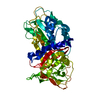
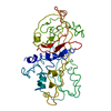
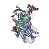
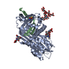
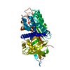
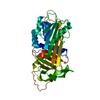
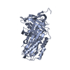
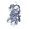
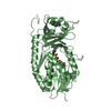
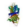
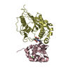
| Title | 2.0 ANGSTROM STRUCTURE OF INTACT ALPHA-1-ANTITRYPSIN: A CANONICAL TEMPLATE FOR ACTIVE SERPINS | |||||||||
 Components Components | ALPHA-1-ANTITRYPSIN Alpha-1 antitrypsin Alpha-1 antitrypsin | |||||||||
 Keywords Keywords | SERINE PROTEASE INHIBITOR / SERPIN / GLYCOPROTEIN / POLYMORPHISM / EMPHYSEMA / DISEASE MUTATION / ACUTE PHASE | |||||||||
| Function / homology |  Function and homology information Function and homology informationCargo concentration in the ER / COPII-mediated vesicle transport / COPII-coated ER to Golgi transport vesicle / endoplasmic reticulum-Golgi intermediate compartment membrane / platelet alpha granule lumen / acute-phase response / Post-translational protein phosphorylation / serine-type endopeptidase inhibitor activity / Regulation of Insulin-like Growth Factor (IGF) transport and uptake by Insulin-like Growth Factor Binding Proteins (IGFBPs) / blood coagulation ...Cargo concentration in the ER / COPII-mediated vesicle transport / COPII-coated ER to Golgi transport vesicle / endoplasmic reticulum-Golgi intermediate compartment membrane / platelet alpha granule lumen / acute-phase response / Post-translational protein phosphorylation / serine-type endopeptidase inhibitor activity / Regulation of Insulin-like Growth Factor (IGF) transport and uptake by Insulin-like Growth Factor Binding Proteins (IGFBPs) / blood coagulation / Platelet degranulation / collagen-containing extracellular matrix / protease binding / ficolin-1-rich granule lumen / endoplasmic reticulum lumen / intracellular membrane-bounded organelle / Neutrophil degranulation / Golgi apparatus / endoplasmic reticulum / extracellular space / extracellular exosome / extracellular region / identical protein bindingSimilarity search - Function | |||||||||
| Biological species |  HOMO SAPIENS (human) HOMO SAPIENS (human) | |||||||||
| Method | X-RAY DIFFRACTION / SYNCHROTRON / MOLECULAR REPLACEMENT / Resolution: 2 Å | |||||||||
 Authors Authors | Elliott, P.R. / Pei, X.Y. / Dafforn, T. / Read, R.J. / Carrell, R.W. / Lomas, D.A. | |||||||||
 Citation Citation | Journal: Protein Sci. / Year: 2000 Title: Topography of a 2.0 A structure of alpha1-antitrypsin reveals targets for rational drug design to prevent conformational disease. Authors: Elliott, P.R. / Pei, X.Y. / Dafforn, T.R. / Lomas, D.A. #1: Journal: J.Mol.Biol. / Year: 1998 Title: Wildtype Alpha1-Antitrypsin is in the Canonical Inhibitory Conformation Authors: Elliott, P.R. / Abrahams, J.-P. / Lomas, D.A. | |||||||||
| History |
|
- Structure visualization
| Structure viewer | Molecule: MolmilJmol/JSmol |
|---|
- Downloads & links
-Download
| PDBx/mmCIF format | 1qlp.cif.gz | 88.3 KB | Display | PDBx/mmCIF format |
|---|---|---|---|---|
| PDB format | pdb1qlp.ent.gz | 66.1 KB | Display | PDB format |
| PDBx/mmJSON format | 1qlp.json.gz | Tree view | PDBx/mmJSON format | |
| Others |  Other downloads Other downloads |
-Validation report
| Arichive directory | https://data.pdbj.org/pub/pdb/validation_reports/ql/1qlpftp://data.pdbj.org/pub/pdb/validation_reports/ql/1qlp | HTTPS FTP |
|---|
-Related structure data
| Related structure data | 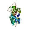 1psiS S: Starting model for refinement |
|---|---|
| Similar structure data |


-Links
 PDBj
PDBj

- Assembly
| Deposited unit | 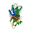
| ||||||||
|---|---|---|---|---|---|---|---|---|---|
| 1 |
| ||||||||
| Unit cell |
|
-Components
| #1: Protein | Alpha-1 antitrypsin / ALPHA-1-PROTEINASE INHIBITOR / ALPHA-1-ANTIPROTEINASE Mass: 44380.441 Da / Num. of mol.: 1 Source method: isolated from a genetically manipulated source Source: (gene. exp.) HOMO SAPIENS (human) / Tissue: HEPATOCYTES / Cell: NEUTROPHILS / Cellular location: CYTOPLASM / Gene: ALPHA-1-ANTITRYPSIN / Organ: LIVER / Plasmid: PTERMAT / Gene (production host): ALPHA-1-ANTITRYPSIN / Production host:  ESCHERICHIA COLI (E. coli) / Strain (production host): BL21(DE3) / References: UniProt: P01009 ESCHERICHIA COLI (E. coli) / Strain (production host): BL21(DE3) / References: UniProt: P01009 |
|---|---|
| #2: Water | ChemComp-HOH / Water Mass: 18.015 Da / Num. of mol.: 106 / Source method: isolated from a natural source / Formula: H2O Mass: 18.015 Da / Num. of mol.: 106 / Source method: isolated from a natural source / Formula: H2O |
-Experimental details
-Experiment
| Experiment | Method: X-RAY DIFFRACTION / Number of used crystals: 1 |
|---|
- Sample preparation
| Crystal | Density Matthews: 2.6 Å3/Da / Density % sol: 37.2 % Description: DATA WERE COLLECTED USING THE OSCILLATION METHOD. | |||||||||||||||||||||||||||||||||||
|---|---|---|---|---|---|---|---|---|---|---|---|---|---|---|---|---|---|---|---|---|---|---|---|---|---|---|---|---|---|---|---|---|---|---|---|---|
| Crystal grow | pH: 6 Details: 24% PEG 4000, 0.2 M SODIUM ACETATE, 0.1M TRIS-HCL PH 6.0, 2MM FESO4.7H20 | |||||||||||||||||||||||||||||||||||
| Crystal grow | *PLUS Temperature: 18 ℃ / Method: vapor diffusion, hanging drop | |||||||||||||||||||||||||||||||||||
| Components of the solutions | *PLUS
|
-Data collection
| Diffraction | Mean temperature: 100 K |
|---|---|
| Diffraction source | Source: SYNCHROTRON / Site: SRS  / Beamline: PX9.6 / Wavelength: 0.87 / Beamline: PX9.6 / Wavelength: 0.87 |
| Detector | Type: MARRESEARCH / Detector: CCD / Date: Sep 9, 1998 / Details: MIRRORS |
| Radiation | Monochromator: BENT CYLINDRICAL GE(111) / Protocol: SINGLE WAVELENGTH / Monochromatic (M) / Laue (L): M / Scattering type: x-ray |
| Radiation wavelength | Wavelength: 0.87 Å / Relative weight: 1 |
| Reflection | Resolution: 2→25 Å / Num. obs: 25057 / % possible obs: 93.6 % / Observed criterion σ(I): 2 / Redundancy: 3.3 % / Biso Wilson estimate: 29.41 Å2 / Rmerge(I) obs: 0.079 / Rsym value: 0.067 / Net I/σ(I): 5.9 |
| Reflection shell | Resolution: 2→2.05 Å / Redundancy: 2.4 % / Rmerge(I) obs: 0.514 / Mean I/σ(I) obs: 1.6 / Rsym value: 0.415 / % possible all: 95.2 |
| Reflection | *PLUS Num. measured all: 156306 |
| Reflection shell | *PLUS Redundancy: 2.2 % / Rmerge(I) obs: 0.503 / Mean I/σ(I) obs: 1.5 |
- Processing
| Software |
| ||||||||||||||||||||||||||||||||||||||||||||||||||||||||||||||||||||||||||||||||
|---|---|---|---|---|---|---|---|---|---|---|---|---|---|---|---|---|---|---|---|---|---|---|---|---|---|---|---|---|---|---|---|---|---|---|---|---|---|---|---|---|---|---|---|---|---|---|---|---|---|---|---|---|---|---|---|---|---|---|---|---|---|---|---|---|---|---|---|---|---|---|---|---|---|---|---|---|---|---|---|---|---|
| Refinement | Method to determine structure: MOLECULAR REPLACEMENT Starting model: THERMOSTABLE VARIANT ALPHA1-ANTITRYPSIN (PDB ENTRY 1PSI) Resolution: 2→25 Å / Data cutoff high absF: 0 / Isotropic thermal model: RESTRAINED / Cross valid method: FREE R-VALUE / σ(F): 0 Details: THE N-TERMINAL RESIDUES 1-22 WERE NOT OBSERVED IN THE ELECTRON DENSITY MAPS
| ||||||||||||||||||||||||||||||||||||||||||||||||||||||||||||||||||||||||||||||||
| Solvent computation | Solvent model: CNS / Bsol: 215 Å2 / ksol: 1.257 e/Å3 | ||||||||||||||||||||||||||||||||||||||||||||||||||||||||||||||||||||||||||||||||
| Displacement parameters | Biso mean: 38.38 Å2
| ||||||||||||||||||||||||||||||||||||||||||||||||||||||||||||||||||||||||||||||||
| Refine analyze |
| ||||||||||||||||||||||||||||||||||||||||||||||||||||||||||||||||||||||||||||||||
| Refinement step | Cycle: LAST / Resolution: 2→25 Å
| ||||||||||||||||||||||||||||||||||||||||||||||||||||||||||||||||||||||||||||||||
| Refine LS restraints |
| ||||||||||||||||||||||||||||||||||||||||||||||||||||||||||||||||||||||||||||||||
| LS refinement shell | Resolution: 2→2.07 Å / Total num. of bins used: 10
| ||||||||||||||||||||||||||||||||||||||||||||||||||||||||||||||||||||||||||||||||
| Xplor file |
| ||||||||||||||||||||||||||||||||||||||||||||||||||||||||||||||||||||||||||||||||
| Refine LS restraints | *PLUS
|
