 Movie
Movie Controller

[English] 日本語
 Yorodumi
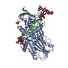
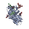
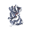
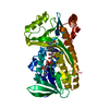
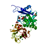
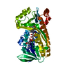
- PDB-8api: THE S VARIANT OF HUMAN ALPHA1-ANTITRYPSIN, STRUCTURE AND IMPLICAT... -
+ Open data

- Basic information
| Entry | Database: PDB / ID: 8api | ||||||||||||
|---|---|---|---|---|---|---|---|---|---|---|---|---|---|
| Title | THE S VARIANT OF HUMAN ALPHA1-ANTITRYPSIN, STRUCTURE AND IMPLICATIONS FOR FUNCTION AND METABOLISM | ||||||||||||
 Components Components | (ALPHA-1 ANTITRYPSIN (CHAIN ...) x 2 | ||||||||||||
 Keywords Keywords |  PROTEINASE INHIBITOR PROTEINASE INHIBITOR | ||||||||||||
| Function / homology |  Function and homology information Function and homology informationCargo concentration in the ER / COPII-mediated vesicle transport / COPII-coated ER to Golgi transport vesicle / endoplasmic reticulum-Golgi intermediate compartment membrane / platelet alpha granule lumen / acute-phase response / Post-translational protein phosphorylation / serine-type endopeptidase inhibitor activity / Regulation of Insulin-like Growth Factor (IGF) transport and uptake by Insulin-like Growth Factor Binding Proteins (IGFBPs) / blood coagulation ...Cargo concentration in the ER / COPII-mediated vesicle transport / COPII-coated ER to Golgi transport vesicle / endoplasmic reticulum-Golgi intermediate compartment membrane / platelet alpha granule lumen / acute-phase response / Post-translational protein phosphorylation / serine-type endopeptidase inhibitor activity / Regulation of Insulin-like Growth Factor (IGF) transport and uptake by Insulin-like Growth Factor Binding Proteins (IGFBPs) / blood coagulation / Platelet degranulation / collagen-containing extracellular matrix / protease binding / ficolin-1-rich granule lumen / endoplasmic reticulum lumen / intracellular membrane-bounded organelle / Neutrophil degranulation / Golgi apparatus / endoplasmic reticulum / extracellular space / extracellular exosome / extracellular region / identical protein bindingSimilarity search - Function | ||||||||||||
| Biological species |  Homo sapiens (human) Homo sapiens (human) | ||||||||||||
| Method | X-RAY DIFFRACTION / Resolution: 3.1 Å | ||||||||||||
 Authors Authors | Loebermann, H. / Tokuoka, R. / Deisenhofer, J. / Huber, R. | ||||||||||||
 Citation Citation | Journal: Protein Eng. / Year: 1989 Title: The S variant of human alpha 1-antitrypsin, structure and implications for function and metabolism. Authors: Engh, R. / Lobermann, H. / Schneider, M. / Wiegand, G. / Huber, R. / Laurell, C.B. #1: Journal: J.Mol.Biol. / Year: 1984Title: Human Alpha1-Proteinase Inhibitor. Crystal Structure Analysis of Two Crystal Modifications, Molecular Model and Preliminary Analysis of the Implications for Function Authors: Loebermann, H. / Tokuoka, R. / Deisenhofer, J. / Huber, R. #2: Journal: Hoppe-Seyler's Z.Physiol.Chem. / Year: 1982Title: Interaction of Human Alpha1-Proteinase Inhibitor with Chymotrypsinogena and Crystallization of a Proteolytically Modified Alpha1-Proteinase Inhibitor Authors: Loebermann, H. / Lottspeich, F. / Bode, W. / Huber, R. #3: Journal: J.Biol.Chem. / Year: 1982Title: The Biosynthesis of Rat Alpha1-Antitrypsin Authors: Carlson, J. / Stenflo, J. #4: Journal: FEBS Lett. / Year: 1981Title: Human Alpha1-Antitrypsin. Carbohydrate Attachment and Sequence Homology Authors: Carrell, R.W. / Jeppsson, J.-O. / Vaughan, L. / Brennan, S.O. / Owen, M.C. / Boswell, D.R. #5: Journal: J.Biol.Chem. / Year: 1980Title: Studies on the Oligosaccharide Chains of Human Alpha1-Protease Inhibitor. II. Structure of Oligosaccharides Authors: Mega, T. / Lujan, E. / Yoshida, A. | ||||||||||||
| History |
|
- Structure visualization
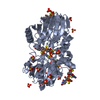
| Structure viewer | Molecule: MolmilJmol/JSmol |
|---|
- Downloads & links
-Download
| PDBx/mmCIF format | 8api.cif.gz | 93.3 KB | Display | PDBx/mmCIF format |
|---|---|---|---|---|
| PDB format | pdb8api.ent.gz | 70.6 KB | Display | PDB format |
| PDBx/mmJSON format | 8api.json.gz | Tree view | PDBx/mmJSON format | |
| Others |  Other downloads Other downloads |
-Validation report
| Arichive directory | https://data.pdbj.org/pub/pdb/validation_reports/ap/8apiftp://data.pdbj.org/pub/pdb/validation_reports/ap/8api | HTTPS FTP |
|---|
-Related structure data


-Links
 PDBj
PDBj


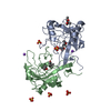
- Assembly
| Deposited unit | 
| ||||||||
|---|---|---|---|---|---|---|---|---|---|
| 1 |
| ||||||||
| Unit cell |
| ||||||||
| Atom site foot note | 1: RESIDUE PRO B 361 IS A CIS PROLINE. 2: AN OCCUPANCY AND B VALUE OF 0.0 INDICATES THAT NO SIGNIFICANT ELECTRON DENSITY WAS FOUND IN THE FINAL FOURIER MAP. |
-Components
-ALPHA-1 ANTITRYPSIN (CHAIN ... , 2 types, 2 molecules AB
| #1: Protein | Mass: 39114.320 Da / Num. of mol.: 1 / Source method: isolated from a natural source / Source: (natural) Homo sapiens (human) / References: UniProt: P01009 |
|---|---|
| #2: Protein/peptide | Mass: 4139.938 Da / Num. of mol.: 1 / Source method: isolated from a natural source / Source: (natural) Homo sapiens (human) / References: UniProt: P01009 |
-Sugars , 2 types, 3 molecules
| #3: Polysaccharide | 2-acetamido-2-deoxy-beta-D-glucopyranose-(1-2)-alpha-D-mannopyranose-(1-6)-[alpha-D-mannopyranose- ...2-acetamido-2-deoxy-beta-D-glucopyranose-(1-2)-alpha-D-mannopyranose-(1-6)-[alpha-D-mannopyranose-(1-3)]alpha-D-mannopyranose-(1-4)-2-acetamido-2-deoxy-beta-D-glucopyranose-(1-4)-2-acetamido-2-deoxy-beta-D-glucopyranose / Mass: 1114.016 Da / Num. of mol.: 1 Source method: isolated from a genetically manipulated source |
|---|---|
| #4: Polysaccharide | / Mass: 424.401 Da / Num. of mol.: 2 Source method: isolated from a genetically manipulated source |
-Non-polymers , 2 types, 160 molecules 

| #5: Chemical | ChemComp-CYS / Cysteine Type: L-peptide linking / Mass: 121.158 Da / Num. of mol.: 1 / Source method: obtained synthetically / Formula: C3H7NO2S Type: L-peptide linking / Mass: 121.158 Da / Num. of mol.: 1 / Source method: obtained synthetically / Formula: C3H7NO2S |
|---|---|
| #6: Water | ChemComp-HOH / WaterMass: 18.015 Da / Num. of mol.: 159 / Source method: isolated from a natural source / Formula: H2O |
-Details
| Compound details | IN THIS ENTRY CHAIN IDENTIFIER *C* IS USED FOR A CYSTEINE RESIDUE WHICH FORMS A DISULFIDE BOND WITH ...IN THIS ENTRY CHAIN IDENTIFIER |
|---|---|
| Nonpolymer details | THE CARBOHYDRATE CHAINS PRESENTED AT THE END OF THIS ENTRY ARE NOT COMPLETE. SOME OF THE SUGAR ...THE CARBOHYDRA |
-Experimental details
-Experiment
| Experiment | Method: X-RAY DIFFRACTION |
|---|
- Sample preparation
| Crystal | Density Matthews: 5.17 Å3/Da / Density % sol: 76.21 % | ||||||||||||||||||||
|---|---|---|---|---|---|---|---|---|---|---|---|---|---|---|---|---|---|---|---|---|---|
| Crystal grow | *PLUS Temperature: 4 ℃ / pH: 8 / Method: vapor diffusionDetails: Loebermann, H., (1982) Hoppe-Seyler'S Z.Physiol. Chem., 363, 1377. | ||||||||||||||||||||
| Components of the solutions | *PLUS
|
-Data collection
| Reflection | *PLUS Highest resolution: 3.1 Å / Num. obs: 15607 / % possible obs: 90 % / Biso Wilson estimate: 276 Å2 |
|---|---|
| Reflection shell | *PLUS Num. possible: 62 |
- Processing
| Software | Name: EREF / Classification: refinement | ||||||||||||||||||||||||||||||||||||||||||||||||||||||||||||
|---|---|---|---|---|---|---|---|---|---|---|---|---|---|---|---|---|---|---|---|---|---|---|---|---|---|---|---|---|---|---|---|---|---|---|---|---|---|---|---|---|---|---|---|---|---|---|---|---|---|---|---|---|---|---|---|---|---|---|---|---|---|
| Refinement | Rfactor Rwork: 0.215 / Highest resolution: 3.1 Å Details: AN OCCUPANCY AND B VALUE OF 0.0 INDICATES THAT NO SIGNIFICANT ELECTRON DENSITY WAS FOUND IN THE FINAL FOURIER MAP. ATOMS WITH THERMAL FACTORS WHICH CALCULATE LESS THAN 6.00 ARE ASSIGNED THIS ...Details: AN OCCUPANCY AND B VALUE OF 0.0 INDICATES THAT NO SIGNIFICANT ELECTRON DENSITY WAS FOUND IN THE FINAL FOURIER MAP. ATOMS WITH THERMAL FACTORS WHICH CALCULATE LESS THAN 6.00 ARE ASSIGNED THIS VALUE. THIS IS THE LOWEST VALUE ALLOWED BY THE REFINEMENT PROGRAM. | ||||||||||||||||||||||||||||||||||||||||||||||||||||||||||||
| Refinement step | Cycle: LAST / Highest resolution: 3.1 Å
| ||||||||||||||||||||||||||||||||||||||||||||||||||||||||||||
| Refine LS restraints |
| ||||||||||||||||||||||||||||||||||||||||||||||||||||||||||||
| Refinement | *PLUS Highest resolution: 3.1 Å / Lowest resolution: 9999 Å / Num. reflection obs: 15607 / Rfactor obs: 0.215 | ||||||||||||||||||||||||||||||||||||||||||||||||||||||||||||
| Solvent computation | *PLUS | ||||||||||||||||||||||||||||||||||||||||||||||||||||||||||||
| Displacement parameters | *PLUS | ||||||||||||||||||||||||||||||||||||||||||||||||||||||||||||
| Refine LS restraints | *PLUS Type: o_angle_d |
