 Movie
Movie Controller


+ Open data

- Basic information
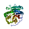
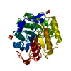
| Entry | Database: PDB / ID: 1qfc | ||||||
|---|---|---|---|---|---|---|---|
| Title | STRUCTURE OF RAT PURPLE ACID PHOSPHATASE | ||||||
 Components Components | PROTEIN (PURPLE ACID PHOSPHATASE) | ||||||
 Keywords Keywords |  HYDROLASE / METAL PHOSPHATASE HYDROLASE / METAL PHOSPHATASE | ||||||
| Function / homology |  Function and homology information Function and homology informationVitamin B2 (riboflavin) metabolism / negative regulation of superoxide anion generation / response to macrophage colony-stimulating factor / negative regulation of macrophage cytokine production / response to L-ascorbic acid / acid phosphatase / acid phosphatase activity / cellular response to zinc ion starvation / negative regulation of nitric oxide biosynthetic process / negative regulation of interleukin-12 production ...Vitamin B2 (riboflavin) metabolism / negative regulation of superoxide anion generation / response to macrophage colony-stimulating factor / negative regulation of macrophage cytokine production / response to L-ascorbic acid / acid phosphatase / acid phosphatase activity / cellular response to zinc ion starvation / negative regulation of nitric oxide biosynthetic process / negative regulation of interleukin-12 production / negative regulation of cell adhesion / response to cholesterol / superoxide anion generation / bone morphogenesis / negative regulation of interleukin-1 beta production / response to zinc ion / negative regulation of tumor necrosis factor production / multicellular organismal response to stress / response to mechanical stimulus / dephosphorylation / bone resorption / nitric oxide biosynthetic process / ossification / ferric iron binding / osteoclast differentiation / response to cytokine / ferrous iron binding / response to insulin / response to organic cyclic compound / negative regulation of inflammatory response / response to ethanol / response to lipopolysaccharide / lysosome / hydrolase activity / positive regulation of cell migration / defense response to Gram-positive bacterium / extracellular spaceSimilarity search - Function | ||||||
| Biological species |  Rattus norvegicus (Norway rat) Rattus norvegicus (Norway rat) | ||||||
| Method | X-RAY DIFFRACTION / MAD / Resolution: 2.7 Å | ||||||
 Authors Authors | Uppenberg, J. / Lindqvist, F. / Svensson, C. / Ek-Rylander, B. / Andersson, G. | ||||||
 Citation Citation | Journal: J.Mol.Biol. / Year: 1999 Title: Crystal structure of a mammalian purple acid phosphatase. Authors: Uppenberg, J. / Lindqvist, F. / Svensson, C. / Ek-Rylander, B. / Andersson, G. | ||||||
| History |
|
- Structure visualization
| Structure viewer | Molecule: MolmilJmol/JSmol |
|---|
- Downloads & links
-Download
| PDBx/mmCIF format | 1qfc.cif.gz | 70.6 KB | Display | PDBx/mmCIF format |
|---|---|---|---|---|
| PDB format | pdb1qfc.ent.gz | 52.1 KB | Display | PDB format |
| PDBx/mmJSON format | 1qfc.json.gz | Tree view | PDBx/mmJSON format | |
| Others |  Other downloads Other downloads |
-Validation report
| Arichive directory | https://data.pdbj.org/pub/pdb/validation_reports/qf/1qfcftp://data.pdbj.org/pub/pdb/validation_reports/qf/1qfc | HTTPS FTP |
|---|
-Related structure data
| Similar structure data |
|---|


-Links
 PDBj
PDBj
- Assembly
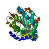
| Deposited unit | 
| ||||||||
|---|---|---|---|---|---|---|---|---|---|
| 1 |
| ||||||||
| Unit cell |
|
-Components
| #1: Protein | Mass: 34392.988 Da / Num. of mol.: 1 Source method: isolated from a genetically manipulated source Source: (gene. exp.) Rattus norvegicus (Norway rat) / Production host:   Spodoptera frugiperda (fall armyworm) / References: UniProt: P29288, acid phosphatase Spodoptera frugiperda (fall armyworm) / References: UniProt: P29288, acid phosphatase | ||
|---|---|---|---|
| #2: Sugar | ChemComp-NAG / N-Acetylglucosamine  Type: D-saccharide, beta linking / Mass: 221.208 Da / Num. of mol.: 1 Type: D-saccharide, beta linking / Mass: 221.208 Da / Num. of mol.: 1Source method: isolated from a genetically manipulated source Formula: C8H15NO6 | ||
| #3: Chemical | Iron  Mass: 55.845 Da / Num. of mol.: 2 / Source method: obtained synthetically / Formula: Fe Mass: 55.845 Da / Num. of mol.: 2 / Source method: obtained synthetically / Formula: Fe#4: Chemical | ChemComp-PO4 / | Phosphate  Mass: 94.971 Da / Num. of mol.: 1 / Source method: obtained synthetically / Formula: PO4 Mass: 94.971 Da / Num. of mol.: 1 / Source method: obtained synthetically / Formula: PO4 |
-Experimental details
-Experiment
| Experiment | Method: X-RAY DIFFRACTION / Number of used crystals: 1 |
|---|
- Sample preparation
| Crystal | Density Matthews: 3.1 Å3/Da / Density % sol: 61 % | ||||||||||||||||||||||||||||||
|---|---|---|---|---|---|---|---|---|---|---|---|---|---|---|---|---|---|---|---|---|---|---|---|---|---|---|---|---|---|---|---|
| Crystal grow | pH: 7 / Details: pH 7.0 | ||||||||||||||||||||||||||||||
| Crystal | *PLUS | ||||||||||||||||||||||||||||||
| Crystal grow | *PLUS Temperature: 18 ℃ / pH: 6.4 / Method: vapor diffusion, hanging drop | ||||||||||||||||||||||||||||||
| Components of the solutions | *PLUS
|
-Data collection
| Diffraction | Mean temperature: 100 K |
|---|---|
| Diffraction source | Source: ROTATING ANODE / Type: RIGAKU RU200 / Wavelength: 1.5418 |
| Detector | Type: RIGAKU RAXIS IV / Detector: IMAGE PLATE / Date: Jan 15, 1998 / Details: YALE MIRRORS |
| Radiation | Protocol: SINGLE WAVELENGTH / Monochromatic (M) / Laue (L): M / Scattering type: x-ray |
| Radiation wavelength | Wavelength: 1.5418 Å / Relative weight: 1 |
| Reflection | Resolution: 2.7→40 Å / Num. obs: 10486 / % possible obs: 90.5 % / Redundancy: 3.9 % / Biso Wilson estimate: 57.9 Å2 / Rsym value: 0.107 / Net I/σ(I): 8 |
| Reflection shell | Resolution: 2.7→2.8 Å / Redundancy: 3.9 % / Mean I/σ(I) obs: 2 / Rsym value: 0.52 / % possible all: 70.1 |
| Reflection | *PLUS Rmerge(I) obs: 0.107 |
- Processing
| Software |
| ||||||||||||||||||||||||||||||||||||||||||||||||||||||||||||||||||||||||||||||||
|---|---|---|---|---|---|---|---|---|---|---|---|---|---|---|---|---|---|---|---|---|---|---|---|---|---|---|---|---|---|---|---|---|---|---|---|---|---|---|---|---|---|---|---|---|---|---|---|---|---|---|---|---|---|---|---|---|---|---|---|---|---|---|---|---|---|---|---|---|---|---|---|---|---|---|---|---|---|---|---|---|---|
| Refinement | Method to determine structure: MAD / Resolution: 2.7→15 Å / Rfactor Rfree error: 0.009 / Data cutoff high rms absF: 1607036.24 / Isotropic thermal model: RESTRAINED / Cross valid method: THROUGHOUT / σ(F): 0
| ||||||||||||||||||||||||||||||||||||||||||||||||||||||||||||||||||||||||||||||||
| Solvent computation | Solvent model: FLAT MODEL / Bsol: 42.13 Å2 / ksol: 0.328 e/Å3 | ||||||||||||||||||||||||||||||||||||||||||||||||||||||||||||||||||||||||||||||||
| Displacement parameters | Biso mean: 57.3 Å2
| ||||||||||||||||||||||||||||||||||||||||||||||||||||||||||||||||||||||||||||||||
| Refine analyze |
| ||||||||||||||||||||||||||||||||||||||||||||||||||||||||||||||||||||||||||||||||
| Refinement step | Cycle: LAST / Resolution: 2.7→15 Å
| ||||||||||||||||||||||||||||||||||||||||||||||||||||||||||||||||||||||||||||||||
| Refine LS restraints |
| ||||||||||||||||||||||||||||||||||||||||||||||||||||||||||||||||||||||||||||||||
| LS refinement shell | Resolution: 2.7→2.87 Å / Rfactor Rfree error: 0.033 / Total num. of bins used: 6
| ||||||||||||||||||||||||||||||||||||||||||||||||||||||||||||||||||||||||||||||||
| Xplor file |
| ||||||||||||||||||||||||||||||||||||||||||||||||||||||||||||||||||||||||||||||||
| Software | *PLUS Name: CNS / Version: 0.4 / Classification: refinement | ||||||||||||||||||||||||||||||||||||||||||||||||||||||||||||||||||||||||||||||||
| Refinement | *PLUS σ(F): 0 / % reflection Rfree: 10.3 % / Rfactor obs: 0.234 | ||||||||||||||||||||||||||||||||||||||||||||||||||||||||||||||||||||||||||||||||
| Solvent computation | *PLUS | ||||||||||||||||||||||||||||||||||||||||||||||||||||||||||||||||||||||||||||||||
| Displacement parameters | *PLUS Biso mean: 57.3 Å2 | ||||||||||||||||||||||||||||||||||||||||||||||||||||||||||||||||||||||||||||||||
| Refine LS restraints | *PLUS
| ||||||||||||||||||||||||||||||||||||||||||||||||||||||||||||||||||||||||||||||||
| LS refinement shell | *PLUS Rfactor Rfree: 0.382 / % reflection Rfree: 10.8 % / Rfactor Rwork: 0.331 |
