 Movie
Movie Controller

[English] 日本語
 Yorodumi
- PDB-3pzv: C2 crystal form of the endo-1,4-beta-glucanase from Bacillus subt... -
+ Open data

- Basic information
| Entry | Database: PDB / ID: 3pzv | ||||||
|---|---|---|---|---|---|---|---|
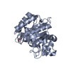
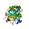
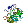
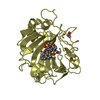
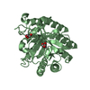
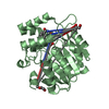
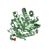
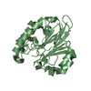
| Title | C2 crystal form of the endo-1,4-beta-glucanase from Bacillus subtilis 168 | ||||||
 Components Components | Endoglucanase Cellulase Cellulase | ||||||
 Keywords Keywords | HYDROLASE / alpha/beta barrel / glycosyl hydrolase / cellulose binding | ||||||
| Function / homology |  Function and homology information Function and homology informationglucan catabolic process / cellulose binding / beta-glucosidase activity / cellulase / cellulase activity / cellulose catabolic process / cell surface / extracellular regionSimilarity search - Function | ||||||
| Biological species |  Bacillus subtilis subsp. subtilis (bacteria) Bacillus subtilis subsp. subtilis (bacteria) | ||||||
| Method | X-RAY DIFFRACTION / SYNCHROTRON / MOLECULAR REPLACEMENT / Resolution: 2.867 Å | ||||||
 Authors Authors | Santos, C.R. / Paiva, J.H. / Akao, P.K. / Meza, A.N. / Silva, J.C. / Squina, F.M. / Ward, R.J. / Ruller, R. / Murakami, M.T. | ||||||
 Citation Citation | Journal: Biochem.J. / Year: 2012 Title: Dissecting structure-function-stability relationships of a thermostable GH5-CBM3 cellulase from Bacillus subtilis 168. Authors: Santos, C.R. / Paiva, J.H. / Sforca, M.L. / Neves, J.L. / Navarro, R.Z. / Cota, J. / Akao, P.K. / Hoffmam, Z.B. / Meza, A.N. / Smetana, J.H. / Nogueira, M.L. / Polikarpov, I. / Xavier-Neto, ...Authors: Santos, C.R. / Paiva, J.H. / Sforca, M.L. / Neves, J.L. / Navarro, R.Z. / Cota, J. / Akao, P.K. / Hoffmam, Z.B. / Meza, A.N. / Smetana, J.H. / Nogueira, M.L. / Polikarpov, I. / Xavier-Neto, J. / Squina, F.M. / Ward, R.J. / Ruller, R. / Zeri, A.C. / Murakami, M.T. | ||||||
| History |
|
- Structure visualization
| Structure viewer | Molecule: MolmilJmol/JSmol |
|---|
- Downloads & links
-Download
| PDBx/mmCIF format | 3pzv.cif.gz | 236.6 KB | Display | PDBx/mmCIF format |
|---|---|---|---|---|
| PDB format | pdb3pzv.ent.gz | 191.9 KB | Display | PDB format |
| PDBx/mmJSON format | 3pzv.json.gz | Tree view | PDBx/mmJSON format | |
| Others |  Other downloads Other downloads |
-Validation report
| Arichive directory | https://data.pdbj.org/pub/pdb/validation_reports/pz/3pzvftp://data.pdbj.org/pub/pdb/validation_reports/pz/3pzv | HTTPS FTP |
|---|
-Related structure data


-Links
 PDBj
PDBj
- Assembly
| Deposited unit | 
| ||||||||
|---|---|---|---|---|---|---|---|---|---|
| 1 | 
| ||||||||
| 2 | 
| ||||||||
| 3 | 
| ||||||||
| 4 | 
| ||||||||
| Unit cell |
|
-Components
| #1: Protein | Cellulase / Carboxymethyl-cellulase / CMCase / Cellulase / Endo-1 / 4-beta-glucanase Mass: 36148.203 Da / Num. of mol.: 4 / Fragment: catalytic domain, Unp residues 27-332 Source method: isolated from a genetically manipulated source Source: (gene. exp.) Bacillus subtilis subsp. subtilis (bacteria)Strain: 168 / Gene: eglS, bglC, gld, BSU18130 / Production host: Escherichia coli (E. coli) / References: UniProt: P10475, cellulase#2: Water | ChemComp-HOH / | Water Mass: 18.015 Da / Num. of mol.: 30 / Source method: isolated from a natural source / Formula: H2O Mass: 18.015 Da / Num. of mol.: 30 / Source method: isolated from a natural source / Formula: H2O |
|---|
-Experimental details
-Experiment
| Experiment | Method: X-RAY DIFFRACTION / Number of used crystals: 1 |
|---|
- Sample preparation
| Crystal | Density Matthews: 2.41 Å3/Da / Density % sol: 49 % |
|---|---|
| Crystal grow | Temperature: 291 K / Method: vapor diffusion, sitting drop / pH: 6.5 Details: 20% PEG3350 200 mM sodium Nitrate, pH 6.5, VAPOR DIFFUSION, SITTING DROP, temperature 291K |
-Data collection
| Diffraction | Mean temperature: 100 K |
|---|---|
| Diffraction source | Source: SYNCHROTRON / Site: LNLS  / Beamline: W01B-MX2 / Wavelength: 1.4586 Å / Beamline: W01B-MX2 / Wavelength: 1.4586 Å |
| Detector | Type: MARMOSAIC 225 mm CCD / Detector: CCD / Date: Sep 9, 2010 |
| Radiation | Protocol: SINGLE WAVELENGTH / Monochromatic (M) / Laue (L): M / Scattering type: x-ray |
| Radiation wavelength | Wavelength: 1.4586 Å / Relative weight: 1 |
| Reflection | Resolution: 2.867→41.5 Å / Num. all: 57202 / Num. obs: 30407 / % possible obs: 97.1 % / Observed criterion σ(I): 2 |
| Reflection shell | Resolution: 2.9→3 Å / % possible all: 83.7 |
- Processing
| Software |
| |||||||||||||||||||||||||||||||||||||||||||||||||||||||||||||||||||||||||||||
|---|---|---|---|---|---|---|---|---|---|---|---|---|---|---|---|---|---|---|---|---|---|---|---|---|---|---|---|---|---|---|---|---|---|---|---|---|---|---|---|---|---|---|---|---|---|---|---|---|---|---|---|---|---|---|---|---|---|---|---|---|---|---|---|---|---|---|---|---|---|---|---|---|---|---|---|---|---|---|
| Refinement | Method to determine structure: MOLECULAR REPLACEMENT / Resolution: 2.867→41.464 Å / SU ML: 0.41 / σ(F): 0 / Stereochemistry target values: ML
| |||||||||||||||||||||||||||||||||||||||||||||||||||||||||||||||||||||||||||||
| Solvent computation | Shrinkage radii: 0.95 Å / VDW probe radii: 1.2 Å / Solvent model: FLAT BULK SOLVENT MODEL / Bsol: 38.056 Å2 / ksol: 0.288 e/Å3 | |||||||||||||||||||||||||||||||||||||||||||||||||||||||||||||||||||||||||||||
| Displacement parameters |
| |||||||||||||||||||||||||||||||||||||||||||||||||||||||||||||||||||||||||||||
| Refinement step | Cycle: LAST / Resolution: 2.867→41.464 Å
| |||||||||||||||||||||||||||||||||||||||||||||||||||||||||||||||||||||||||||||
| Refine LS restraints |
| |||||||||||||||||||||||||||||||||||||||||||||||||||||||||||||||||||||||||||||
| LS refinement shell |
|
