 Movie
Movie Controller

[English] 日本語
 Yorodumi
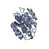
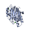
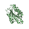
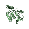
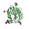
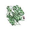
- PDB-1qcy: THE CRYSTAL STRUCTURE OF THE I-DOMAIN OF HUMAN INTEGRIN ALPHA1BETA1 -
+ Open data

- Basic information
| Entry | Database: PDB / ID: 1qcy | ||||||
|---|---|---|---|---|---|---|---|
| Title | THE CRYSTAL STRUCTURE OF THE I-DOMAIN OF HUMAN INTEGRIN ALPHA1BETA1 | ||||||
 Components Components | I-DOMAIN OF INTEGRIN ALPHA1BETA1 | ||||||
 Keywords Keywords |  CELL ADHESION / DINUCLEOTIDE BINDING FOLD / ROSSMANN FOLD CELL ADHESION / DINUCLEOTIDE BINDING FOLD / ROSSMANN FOLD | ||||||
| Function / homology |  Function and homology information Function and homology informationcellular extravasation / integrin alpha1-beta1 complex / collagen binding involved in cell-matrix adhesion / Other semaphorin interactions / CHL1 interactions / Laminin interactions / basal part of cell / Platelet Adhesion to exposed collagen / integrin complex / cell adhesion mediated by integrin ...cellular extravasation / integrin alpha1-beta1 complex / collagen binding involved in cell-matrix adhesion / Other semaphorin interactions / CHL1 interactions / Laminin interactions / basal part of cell / Platelet Adhesion to exposed collagen / integrin complex / cell adhesion mediated by integrin / negative regulation of epidermal growth factor receptor signaling pathway / positive regulation of phosphoprotein phosphatase activity / Smooth Muscle Contraction / Integrin cell surface interactions / collagen binding / cell-matrix adhesion / neutrophil chemotaxis / neuron projection morphogenesis / acrosomal vesicle / integrin-mediated signaling pathway / cell-cell adhesion / vasodilation / positive regulation of neuron apoptotic process / integrin binding / perikaryon / protein phosphatase binding / positive regulation of MAPK cascade / negative regulation of cell population proliferation / external side of plasma membrane / focal adhesion / cell surface / extracellular exosome / membrane / metal ion binding / plasma membraneSimilarity search - Function | ||||||
| Biological species |  Homo sapiens (human) Homo sapiens (human) | ||||||
| Method | X-RAY DIFFRACTION / Resolution: 2.3 Å | ||||||
 Authors Authors | Kankare, J.A. / Salminen, T.A. / Nymalm, Y. / Kaepylae, J. / Heino, J. / Johnson, M.S. | ||||||
 Citation Citation | Journal: J.Biol.Chem. / Year: 2004 Title: Jararhagin-derived RKKH Peptides Induce Structural Changes in a1I Domain of Human Integrin a1b1 Authors: Nymalm, Y. / Puranen, J.S. / Nyholm, T.K.M. / Kaepylae, J. / Kidron, H. / Pentikaeinen, O.T. / Airenne, T.T. / Heino, J. / Slotte, J.P. / Johnson, M.S. / Salminen, T.A. | ||||||
| History |
|
- Structure visualization
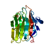
| Structure viewer | Molecule: MolmilJmol/JSmol |
|---|
- Downloads & links
-Download
| PDBx/mmCIF format | 1qcy.cif.gz | 52.7 KB | Display | PDBx/mmCIF format |
|---|---|---|---|---|
| PDB format | pdb1qcy.ent.gz | 36.7 KB | Display | PDB format |
| PDBx/mmJSON format | 1qcy.json.gz | Tree view | PDBx/mmJSON format | |
| Others |  Other downloads Other downloads |
-Validation report
| Arichive directory | https://data.pdbj.org/pub/pdb/validation_reports/qc/1qcyftp://data.pdbj.org/pub/pdb/validation_reports/qc/1qcy | HTTPS FTP |
|---|
-Related structure data

-Links
 PDBj
PDBj


- Assembly
| Deposited unit | 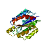
| ||||||||||
|---|---|---|---|---|---|---|---|---|---|---|---|
| 1 |
| ||||||||||
| Unit cell |
| ||||||||||
| Details | I-domain is a fragment of alpha1 subunit of the large integrin heterodimer |
-Components
| #1: Protein | Mass: 21546.402 Da / Num. of mol.: 1 / Fragment: FRAGMENT, I-DOMAIN OF HUMAN INTEGRIN A1B1 / Mutation: K170E Source method: isolated from a genetically manipulated source Source: (gene. exp.) Homo sapiens (human) / Production host:  Escherichia coli (E. coli)Keywords: THE SEQUENCE IS DIFFERENT IN SWISS-PROT, NO ELECTRON SIDE-CHAIN DENSITY WAS EVER SEEN FOR RESIDUE R218, LEFT AS AN ALANINE IN THE STRUCTURE Escherichia coli (E. coli)Keywords: THE SEQUENCE IS DIFFERENT IN SWISS-PROT, NO ELECTRON SIDE-CHAIN DENSITY WAS EVER SEEN FOR RESIDUE R218, LEFT AS AN ALANINE IN THE STRUCTUREReferences: UniProt: P56199 |
|---|---|
| #2: Chemical | ChemComp-MG /   Mass: 24.305 Da / Num. of mol.: 1 / Source method: obtained synthetically / Formula: Mg Mass: 24.305 Da / Num. of mol.: 1 / Source method: obtained synthetically / Formula: Mg |
| #3: Water | ChemComp-HOH / Water Mass: 18.015 Da / Num. of mol.: 94 / Source method: isolated from a natural source / Formula: H2O Mass: 18.015 Da / Num. of mol.: 94 / Source method: isolated from a natural source / Formula: H2O |
-Experimental details
-Experiment
| Experiment | Method: X-RAY DIFFRACTION / Number of used crystals: 1 |
|---|
- Sample preparation
| Crystal | Density Matthews: 2.64 Å3/Da / Density % sol: 53.39 % |
|---|---|
| Crystal grow | Temperature: 298 K / Method: vapor diffusion, sitting drop / pH: 8.5 Details: PEG 4000, Na-acetate, glycerol, pH 8.5, VAPOR DIFFUSION, SITTING DROP, temperature 298.0K |
| Crystal grow | *PLUS Method: unknown |
-Data collection
| Diffraction | Mean temperature: 100 K |
|---|---|
| Diffraction source | Source: ROTATING ANODE / Type: RIGAKU RU200 / Wavelength: 1.5418 |
| Detector | Type: RIGAKU RAXIS IIC / Detector: IMAGE PLATE / Date: Sep 10, 1998 |
| Radiation | Protocol: SINGLE WAVELENGTH / Monochromatic (M) / Laue (L): M / Scattering type: x-ray |
| Radiation wavelength | Wavelength: 1.5418 Å / Relative weight: 1 |
| Reflection | Resolution: 2.3→500 Å / Num. obs: 9471 / % possible obs: 94.8 % / Observed criterion σ(F): 0 / Observed criterion σ(I): 0 / Rmerge(I) obs: 0.058 |
| Reflection shell | Resolution: 2.3→2.38 Å / Num. unique all: 796 / % possible all: 90.9 |
- Processing
| Software |
| ||||||||||||||||||||||||||||||||||||||||||||||||||||||||||||||||||||||||||||||||
|---|---|---|---|---|---|---|---|---|---|---|---|---|---|---|---|---|---|---|---|---|---|---|---|---|---|---|---|---|---|---|---|---|---|---|---|---|---|---|---|---|---|---|---|---|---|---|---|---|---|---|---|---|---|---|---|---|---|---|---|---|---|---|---|---|---|---|---|---|---|---|---|---|---|---|---|---|---|---|---|---|---|
| Refinement | Resolution: 2.3→500 Å / Rfactor Rfree error: 0.008 / Data cutoff high absF: 374683.35 / Data cutoff low absF: 0 / Isotropic thermal model: RESTRAINED / Cross valid method: THROUGHOUT / σ(F): 0
| ||||||||||||||||||||||||||||||||||||||||||||||||||||||||||||||||||||||||||||||||
| Solvent computation | Solvent model: FLAT MODEL / Bsol: 29.22 Å2 / ksol: 0.34 e/Å3 | ||||||||||||||||||||||||||||||||||||||||||||||||||||||||||||||||||||||||||||||||
| Displacement parameters | Biso mean: 24.8 Å2
| ||||||||||||||||||||||||||||||||||||||||||||||||||||||||||||||||||||||||||||||||
| Refine analyze |
| ||||||||||||||||||||||||||||||||||||||||||||||||||||||||||||||||||||||||||||||||
| Refinement step | Cycle: LAST / Resolution: 2.3→500 Å
| ||||||||||||||||||||||||||||||||||||||||||||||||||||||||||||||||||||||||||||||||
| Refine LS restraints |
| ||||||||||||||||||||||||||||||||||||||||||||||||||||||||||||||||||||||||||||||||
| LS refinement shell | Resolution: 2.3→2.38 Å / Rfactor Rfree error: 0.03 / Total num. of bins used: 10
| ||||||||||||||||||||||||||||||||||||||||||||||||||||||||||||||||||||||||||||||||
| Xplor file |
|
