 Movie
Movie Controller


+ Open data

- Basic information
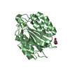
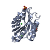
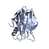
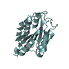
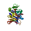
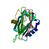
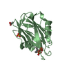
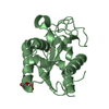
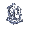
| Entry | Database: PDB / ID: 1ck4 | ||||||
|---|---|---|---|---|---|---|---|
| Title | CRYSTAL STRUCTURE OF RAT A1B1 INTEGRIN I-DOMAIN. | ||||||
 Components Components | INTEGRIN ALPHA-1 | ||||||
 Keywords Keywords | STRUCTURAL PROTEIN / I-DOMAIN / METAL BINDING / COLLAGEN / ADHESION | ||||||
| Function / homology |  Function and homology information Function and homology informationIntegrin cell surface interactions / Smooth Muscle Contraction / cellular extravasation / integrin alpha1-beta1 complex / collagen binding involved in cell-matrix adhesion / basal part of cell / integrin complex / cell adhesion mediated by integrin / negative regulation of epidermal growth factor receptor signaling pathway / positive regulation of phosphoprotein phosphatase activity ...Integrin cell surface interactions / Smooth Muscle Contraction / cellular extravasation / integrin alpha1-beta1 complex / collagen binding involved in cell-matrix adhesion / basal part of cell / integrin complex / cell adhesion mediated by integrin / negative regulation of epidermal growth factor receptor signaling pathway / positive regulation of phosphoprotein phosphatase activity / collagen binding / cell chemotaxis / cell-matrix adhesion / neutrophil chemotaxis / neuron projection morphogenesis / acrosomal vesicle / integrin-mediated signaling pathway / cell-cell adhesion / vasodilation / positive regulation of neuron apoptotic process / integrin binding / perikaryon / protein phosphatase binding / positive regulation of MAPK cascade / cell adhesion / negative regulation of cell population proliferation / external side of plasma membrane / signaling receptor binding / focal adhesion / cell surface / metal ion bindingSimilarity search - Function | ||||||
| Biological species |  Rattus norvegicus (Norway rat) Rattus norvegicus (Norway rat) | ||||||
| Method | X-RAY DIFFRACTION / MOLECULAR REPLACEMENT / Resolution: 2.2 Å | ||||||
 Authors Authors | Nolte, M. / Pepinsky, R.B. / Venyaminov, S.Y. / Koteliansky, V. / Gotwals, P.J. / Karpusas, M. | ||||||
 Citation Citation | Journal: FEBS Lett. / Year: 1999 Title: Crystal structure of the alpha1beta1 integrin I-domain: insights into integrin I-domain function. Authors: Nolte, M. / Pepinsky, R.B. / Venyaminov, S.Y.u. / Koteliansky, V. / Gotwals, P.J. / Karpusas, M. | ||||||
| History |
|
- Structure visualization
| Structure viewer | Molecule: MolmilJmol/JSmol |
|---|
- Downloads & links
-Download
| PDBx/mmCIF format | 1ck4.cif.gz | 106.9 KB | Display | PDBx/mmCIF format |
|---|---|---|---|---|
| PDB format | pdb1ck4.ent.gz | 82.5 KB | Display | PDB format |
| PDBx/mmJSON format | 1ck4.json.gz | Tree view | PDBx/mmJSON format | |
| Others |  Other downloads Other downloads |
-Validation report
| Arichive directory | https://data.pdbj.org/pub/pdb/validation_reports/ck/1ck4ftp://data.pdbj.org/pub/pdb/validation_reports/ck/1ck4 | HTTPS FTP |
|---|
-Related structure data
| Related structure data |  1aoxS S: Starting model for refinement |
|---|---|
| Similar structure data |

-Links
 PDBj
PDBj


- Assembly
| Deposited unit | 
| ||||||||
|---|---|---|---|---|---|---|---|---|---|
| 1 | 
| ||||||||
| 2 | 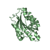
| ||||||||
| Unit cell |
| ||||||||
| Noncrystallographic symmetry (NCS) | NCS oper: (Code: given Matrix: (0.99929, 0.00081, 0.03763), Vector : |
-Components
| #1: Protein | Mass: 22118.053 Da / Num. of mol.: 2 / Fragment: I-DOMAIN Source method: isolated from a genetically manipulated source Source: (gene. exp.) Rattus norvegicus (Norway rat) / Cell line (production host): DH5A / Production host:  Escherichia coli (E. coli) / References: UniProt: P18614 Escherichia coli (E. coli) / References: UniProt: P18614#2: Water | ChemComp-HOH / | Water Mass: 18.015 Da / Num. of mol.: 215 / Source method: isolated from a natural source / Formula: H2O Mass: 18.015 Da / Num. of mol.: 215 / Source method: isolated from a natural source / Formula: H2O |
|---|
-Experimental details
-Experiment
| Experiment | Method: X-RAY DIFFRACTION / Number of used crystals: 1 |
|---|
- Sample preparation
| Crystal | Density Matthews: 4.22 Å3/Da / Density % sol: 45.03 % | |||||||||||||||||||||||||||||||||||
|---|---|---|---|---|---|---|---|---|---|---|---|---|---|---|---|---|---|---|---|---|---|---|---|---|---|---|---|---|---|---|---|---|---|---|---|---|
| Crystal grow | pH: 6.5 / Details: pH 6.5 | |||||||||||||||||||||||||||||||||||
| Crystal grow | *PLUS pH: 7.5 / Method: vapor diffusion | |||||||||||||||||||||||||||||||||||
| Components of the solutions | *PLUS
|
-Data collection
| Diffraction | Mean temperature: 108 K |
|---|---|
| Diffraction source | Source: ROTATING ANODE / Type: RIGAKU RU200 / Wavelength: 1.5418 |
| Detector | Type: RIGAKU / Detector: IMAGE PLATE / Details: MIRRORS |
| Radiation | Monochromator: NI FILTER / Protocol: SINGLE WAVELENGTH / Monochromatic (M) / Laue (L): M / Scattering type: x-ray |
| Radiation wavelength | Wavelength: 1.5418 Å / Relative weight: 1 |
| Reflection | Resolution: 2.2→35 Å / Num. obs: 19238 / % possible obs: 91 % / Observed criterion σ(I): 2 / Redundancy: 12 % / Rmerge(I) obs: 0.056 / Net I/σ(I): 20 |
| Reflection shell | *PLUS Highest resolution: 2.2 Å / Lowest resolution: 2.28 Å / % possible obs: 77.6 % / Mean I/σ(I) obs: 3.14 |
- Processing
| Software |
| ||||||||||||||||||||||||||||||||||||||||||||||||||||||||||||
|---|---|---|---|---|---|---|---|---|---|---|---|---|---|---|---|---|---|---|---|---|---|---|---|---|---|---|---|---|---|---|---|---|---|---|---|---|---|---|---|---|---|---|---|---|---|---|---|---|---|---|---|---|---|---|---|---|---|---|---|---|---|
| Refinement | Method to determine structure: MOLECULAR REPLACEMENT Starting model: PDB ENTRY 1AOX Resolution: 2.2→100 Å / Cross valid method: THROUGHOUT / σ(F): 2 Details: FOR CHAIN A, SIDE CHAINS OF RESIDUES 145, 146, 234 ARE ASSIGNED 0.0 OCCUPANCY DUE TO ABSENCE OF ELECTRON DENSITY FOR THE SIDE CHAINS. FOR CHAIN B, SIDE CHAINS OF RESIDUES 145, 175, 234 ARE ...Details: FOR CHAIN A, SIDE CHAINS OF RESIDUES 145, 146, 234 ARE ASSIGNED 0.0 OCCUPANCY DUE TO ABSENCE OF ELECTRON DENSITY FOR THE SIDE CHAINS. FOR CHAIN B, SIDE CHAINS OF RESIDUES 145, 175, 234 ARE ASSIGNED 0.0 OCCUPANCY DUE TO ABSENCE OF ELECTRON DENSITY FOR THE SIDE CHAINS.
| ||||||||||||||||||||||||||||||||||||||||||||||||||||||||||||
| Refinement step | Cycle: LAST / Resolution: 2.2→100 Å
| ||||||||||||||||||||||||||||||||||||||||||||||||||||||||||||
| Refine LS restraints |
| ||||||||||||||||||||||||||||||||||||||||||||||||||||||||||||
| Software | *PLUS Name: X-PLOR / Version: 3.8 / Classification: refinement | ||||||||||||||||||||||||||||||||||||||||||||||||||||||||||||
| Refinement | *PLUS Lowest resolution: 100 Å / σ(F): 2 / % reflection Rfree: 10 % / Rfactor obs: 0.234 | ||||||||||||||||||||||||||||||||||||||||||||||||||||||||||||
| Solvent computation | *PLUS | ||||||||||||||||||||||||||||||||||||||||||||||||||||||||||||
| Displacement parameters | *PLUS | ||||||||||||||||||||||||||||||||||||||||||||||||||||||||||||
| Refine LS restraints | *PLUS Type: x_angle_deg / Dev ideal: 1.6 |
