 Movie
Movie Controller

[English] 日本語
 Yorodumi
- PDB-1jfu: CRYSTAL STRUCTURE OF THE SOLUBLE DOMAIN OF TLPA FROM BRADYRHIZOBI... -
+ Open data

- Basic information
| Entry | Database: PDB / ID: 1jfu | ||||||
|---|---|---|---|---|---|---|---|
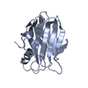
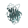
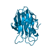
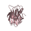
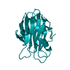
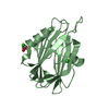
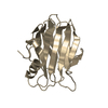
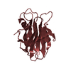
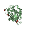
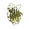
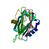
| Title | CRYSTAL STRUCTURE OF THE SOLUBLE DOMAIN OF TLPA FROM BRADYRHIZOBIUM JAPONICUM | ||||||
 Components Components | THIOL:DISULFIDE INTERCHANGE PROTEIN TLPA | ||||||
 Keywords Keywords |  MEMBRANE PROTEIN / THIOREDOXIN-LIKE / DOUBLE DISULFIDE BRIDGE MEMBRANE PROTEIN / THIOREDOXIN-LIKE / DOUBLE DISULFIDE BRIDGE | ||||||
| Function / homology |  Function and homology information Function and homology informationcytochrome complex assembly / disulfide oxidoreductase activity / plasma membraneSimilarity search - Function | ||||||
| Biological species |  Bradyrhizobium japonicum (bacteria) Bradyrhizobium japonicum (bacteria) | ||||||
| Method | X-RAY DIFFRACTION / SIRAS / Resolution: 1.6 Å | ||||||
 Authors Authors | Capitani, G. / Rossmann, R. / Sargent, D.F. / Gruetter, M.G. / Richmond, T.J. / Hennecke, H. | ||||||
 Citation Citation | Journal: J.Mol.Biol. / Year: 2001 Title: Structure of the soluble domain of a membrane-anchored thioredoxin-like protein from Bradyrhizobium japonicum reveals unusual properties. Authors: Capitani, G. / Rossmann, R. / Sargent, D.F. / Grutter, M.G. / Richmond, T.J. / Hennecke, H. | ||||||
| History |
|
- Structure visualization
| Structure viewer | Molecule: MolmilJmol/JSmol |
|---|
- Downloads & links
-Download
| PDBx/mmCIF format | 1jfu.cif.gz | 86.4 KB | Display | PDBx/mmCIF format |
|---|---|---|---|---|
| PDB format | pdb1jfu.ent.gz | 69.7 KB | Display | PDB format |
| PDBx/mmJSON format | 1jfu.json.gz | Tree view | PDBx/mmJSON format | |
| Others |  Other downloads Other downloads |
-Validation report
| Arichive directory | https://data.pdbj.org/pub/pdb/validation_reports/jf/1jfuftp://data.pdbj.org/pub/pdb/validation_reports/jf/1jfu | HTTPS FTP |
|---|
-Related structure data
| Similar structure data |
|---|
-Links
 PDBj
PDBj

- Assembly
| Deposited unit | 
| ||||||||
|---|---|---|---|---|---|---|---|---|---|
| 1 | 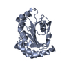
| ||||||||
| 2 | 
| ||||||||
| Unit cell |
|
-Components
| #1: Protein | Mass: 19788.889 Da / Num. of mol.: 2 / Fragment: SOLUBLE DOMAIN OF TLPA (RESIDUES 36-221) Source method: isolated from a genetically manipulated source Source: (gene. exp.) Bradyrhizobium japonicum (bacteria) / Gene: TLPA / Plasmid: PMAL-P / Species (production host): Escherichia coli / Production host: Escherichia coli BL21 (bacteria) / Strain (production host): BL21 / References: UniProt: P43221#2: Water | ChemComp-HOH / | Water Mass: 18.015 Da / Num. of mol.: 555 / Source method: isolated from a natural source / Formula: H2O Mass: 18.015 Da / Num. of mol.: 555 / Source method: isolated from a natural source / Formula: H2O |
|---|
-Experimental details
-Experiment
| Experiment | Method: X-RAY DIFFRACTION / Number of used crystals: 1 |
|---|
- Sample preparation
| Crystal | Density Matthews: 1.99 Å3/Da / Density % sol: 38.28 % | |||||||||||||||||||||||||||||||||||
|---|---|---|---|---|---|---|---|---|---|---|---|---|---|---|---|---|---|---|---|---|---|---|---|---|---|---|---|---|---|---|---|---|---|---|---|---|
| Crystal grow | Temperature: 280 K / Method: vapor diffusion, hanging drop / pH: 9 Details: Bicine, NaCl, PEG550 MME, pH 9.0, VAPOR DIFFUSION, HANGING DROP, temperature 280K | |||||||||||||||||||||||||||||||||||
| Crystal grow | *PLUS Temperature: 7 ℃ | |||||||||||||||||||||||||||||||||||
| Components of the solutions | *PLUS
|
-Data collection
| Diffraction | Mean temperature: 100 K |
|---|---|
| Diffraction source | Source: ROTATING ANODE / Type: RIGAKU RU200 / Wavelength: 1.5418 Å |
| Detector | Type: MARRESEARCH / Detector: IMAGE PLATE / Date: Dec 4, 1999 / Details: OSMIC Confocal Max-Flux |
| Radiation | Monochromator: OSMIC MIRRORS / Protocol: SINGLE WAVELENGTH / Monochromatic (M) / Laue (L): M / Scattering type: x-ray |
| Radiation wavelength | Wavelength: 1.5418 Å / Relative weight: 1 |
| Reflection | Resolution: 1.6→15 Å / Num. obs: 36890 / % possible obs: 86.7 % / Observed criterion σ(I): -3 / Redundancy: 3.5 % / Biso Wilson estimate: 18.6 Å2 / Rmerge(I) obs: 0.062 / Net I/σ(I): 16.2 |
| Reflection shell | Resolution: 1.6→1.67 Å / Redundancy: 2.7 % / Rmerge(I) obs: 0.38 / Mean I/σ(I) obs: 2.5 / Num. unique all: 3346 / % possible all: 64 |
- Processing
| Software |
| |||||||||||||||||||||
|---|---|---|---|---|---|---|---|---|---|---|---|---|---|---|---|---|---|---|---|---|---|---|
| Refinement | Method to determine structure: SIRAS / Resolution: 1.6→15 Å / Isotropic thermal model: ISOTROPIC / Cross valid method: THROUGHOUT / σ(F): 0 / Stereochemistry target values: ENGH & HUBER
| |||||||||||||||||||||
| Displacement parameters | Biso mean: 19.3 Å2
| |||||||||||||||||||||
| Refinement step | Cycle: LAST / Resolution: 1.6→15 Å
| |||||||||||||||||||||
| Refine LS restraints |
| |||||||||||||||||||||
| LS refinement shell |
| |||||||||||||||||||||
| Software | *PLUS Name: REFMAC / Classification: refinement | |||||||||||||||||||||
| Refinement | *PLUS Highest resolution: 1.6 Å / Lowest resolution: 15 Å / σ(F): 0 / % reflection Rfree: 5 % / Rfactor obs: 0.181 | |||||||||||||||||||||
| Solvent computation | *PLUS | |||||||||||||||||||||
| Displacement parameters | *PLUS Biso mean: 19.3 Å2 | |||||||||||||||||||||
| Refine LS restraints | *PLUS
|
