 Movie
Movie Controller

[English] 日本語
 Yorodumi
- PDB-1oxb: Complex between YPD1 and SLN1 response regulator domain in space ... -
+ Open data

- Basic information
| Entry | Database: PDB / ID: 1oxb | ||||||
|---|---|---|---|---|---|---|---|
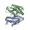
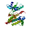
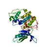
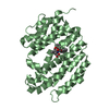
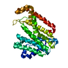
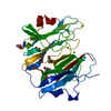
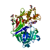
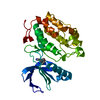
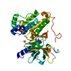
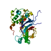
| Title | Complex between YPD1 and SLN1 response regulator domain in space group P2(1)2(1)2(1) | ||||||
 Components Components |
| ||||||
 Keywords Keywords |  SIGNALING PROTEIN / phosphorelay protein / two-component signaling protein / response regulator / HPt domain / Histidine-containing phosphotransfer protein / Ypd1p / Sln1p SIGNALING PROTEIN / phosphorelay protein / two-component signaling protein / response regulator / HPt domain / Histidine-containing phosphotransfer protein / Ypd1p / Sln1p | ||||||
| Function / homology |  Function and homology information Function and homology informationprotein histidine kinase binding / osmosensor activity / transferase activity, transferring phosphorus-containing groups / histidine phosphotransfer kinase activity / osmosensory signaling via phosphorelay pathway / protein histidine kinase activity / histidine kinase / phosphorelay sensor kinase activity / phosphorelay signal transduction system / cell periphery ...protein histidine kinase binding / osmosensor activity / transferase activity, transferring phosphorus-containing groups / histidine phosphotransfer kinase activity / osmosensory signaling via phosphorelay pathway / protein histidine kinase activity / histidine kinase / phosphorelay sensor kinase activity / phosphorelay signal transduction system / cell periphery / phosphorylation / ATP binding / metal ion binding / nucleus / plasma membrane / cytoplasmSimilarity search - Function | ||||||
| Biological species |  Saccharomyces cerevisiae (brewer's yeast) Saccharomyces cerevisiae (brewer's yeast) | ||||||
| Method | X-RAY DIFFRACTION / MOLECULAR REPLACEMENT / Resolution: 2.3 Å | ||||||
 Authors Authors | Xu, Q. / Porter, S.W. / West, A.H. | ||||||
 Citation Citation | Journal: Structure / Year: 2003 Title: The yeast YPD1/SLN1 complex: insights into molecular recognition in two-component signaling systems. Authors: Xu, Q. / Porter, S.W. / West, A.H. #1: Journal: Acta Crystallogr.,Sect.D / Year: 2003Title: Co-Crystallization of the Yeast Phosphorelay Protein YPD1 with the SLN1 Response-Regulator Domain and Preliminary X-ray Diffraction Analysis Authors: Chooback, L. / West, A.H. | ||||||
| History |
|
- Structure visualization
| Structure viewer | Molecule: MolmilJmol/JSmol |
|---|
- Downloads & links
-Download
| PDBx/mmCIF format | 1oxb.cif.gz | 72.2 KB | Display | PDBx/mmCIF format |
|---|---|---|---|---|
| PDB format | pdb1oxb.ent.gz | 52.3 KB | Display | PDB format |
| PDBx/mmJSON format | 1oxb.json.gz | Tree view | PDBx/mmJSON format | |
| Others |  Other downloads Other downloads |
-Validation report
| Arichive directory | https://data.pdbj.org/pub/pdb/validation_reports/ox/1oxbftp://data.pdbj.org/pub/pdb/validation_reports/ox/1oxb | HTTPS FTP |
|---|
-Related structure data
| Related structure data | 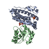 1oxkC  1qspS S: Starting model for refinement C: citing same article ( |
|---|---|
| Similar structure data |
-Links
 PDBj
PDBj


- Assembly
| Deposited unit | 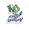
| ||||||||
|---|---|---|---|---|---|---|---|---|---|
| 1 |
| ||||||||
| Unit cell |
|
-Components
| #1: Protein | Mass: 19056.457 Da / Num. of mol.: 1 Source method: isolated from a genetically manipulated source Source: (gene. exp.) Saccharomyces cerevisiae (brewer's yeast)Gene: YPD1 / Plasmid: pUC12 derivative / Production host:  Escherichia coli (E. coli) / Strain (production host): DH5alpha / References: UniProt: Q07688 Escherichia coli (E. coli) / Strain (production host): DH5alpha / References: UniProt: Q07688 |
|---|---|
| #2: Protein | Mass: 15092.516 Da / Num. of mol.: 1 / Fragment: C-terminal residues 1087-1220 Source method: isolated from a genetically manipulated source Source: (gene. exp.) Saccharomyces cerevisiae (brewer's yeast)Gene: SLN1 OR YPD2 OR YIL147C / Plasmid: pCYB2 / Species (production host): Escherichia coli / Production host: Escherichia coli BL21(DE3) (bacteria) / Strain (production host): BL21(DE3)References: UniProt: P39928, Transferases; Transferring phosphorus-containing groups; Phosphotransferases with a nitrogenous group as acceptor |
| #3: Chemical | ChemComp-SO4 / Sulfate  Mass: 96.063 Da / Num. of mol.: 1 / Source method: obtained synthetically / Formula: SO4 Mass: 96.063 Da / Num. of mol.: 1 / Source method: obtained synthetically / Formula: SO4 |
| #4: Water | ChemComp-HOH / Water Mass: 18.015 Da / Num. of mol.: 54 / Source method: isolated from a natural source / Formula: H2O Mass: 18.015 Da / Num. of mol.: 54 / Source method: isolated from a natural source / Formula: H2O |
-Experimental details
-Experiment
| Experiment | Method: X-RAY DIFFRACTION / Number of used crystals: 1 |
|---|
- Sample preparation
| Crystal | Density Matthews: 2.88 Å3/Da / Density % sol: 57.01 % | ||||||||||||||||||||||||
|---|---|---|---|---|---|---|---|---|---|---|---|---|---|---|---|---|---|---|---|---|---|---|---|---|---|
| Crystal grow | Temperature: 296 K / Method: vapor diffusion, hanging drop / pH: 5.3 Details: ammonium sulfate, sodium acetate, BeCl2, NaF, pH 5.3, VAPOR DIFFUSION, HANGING DROP, temperature 296K | ||||||||||||||||||||||||
| Crystal grow | *PLUS Method: vapor diffusion, hanging drop / Details: Choobakc, L., (2003) Acta Cryst., D59, 927. | ||||||||||||||||||||||||
| Components of the solutions | *PLUS
|
-Data collection
| Diffraction | Mean temperature: 100 K |
|---|---|
| Diffraction source | Source: ROTATING ANODE / Type: RIGAKU RUH3R / Wavelength: 1.5418 Å |
| Detector | Type: RIGAKU RAXIS IV / Detector: IMAGE PLATE / Details: osmic confocal mirrors |
| Radiation | Protocol: SINGLE WAVELENGTH / Monochromatic (M) / Laue (L): M / Scattering type: x-ray |
| Radiation wavelength | Wavelength: 1.5418 Å / Relative weight: 1 |
| Reflection | Resolution: 2.3→30 Å / Num. all: 17288 / Num. obs: 17288 / % possible obs: 98.3 % / Observed criterion σ(F): 0 / Observed criterion σ(I): 0 / Redundancy: 4.9 % / Biso Wilson estimate: 28.3 Å2 / Rmerge(I) obs: 0.05 / Net I/σ(I): 19 |
| Reflection shell | Resolution: 2.3→2.44 Å / % possible all: 94.5 |
| Reflection | *PLUS Lowest resolution: 30 Å / Num. obs: 17319 / Num. measured all: 84027 |
| Reflection shell | *PLUS % possible obs: 94.5 % / Rmerge(I) obs: 0.239 / Mean I/σ(I) obs: 6.5 |
- Processing
| Software |
| |||||||||||||||||||||||||
|---|---|---|---|---|---|---|---|---|---|---|---|---|---|---|---|---|---|---|---|---|---|---|---|---|---|---|
| Refinement | Method to determine structure: MOLECULAR REPLACEMENT Starting model: 1QSP (YPD1) Resolution: 2.3→30.16 Å / Rfactor Rfree error: 0.005 / Data cutoff high absF: 1933271.52 / Data cutoff high rms absF: 1933271.52 / Data cutoff low absF: 0 / Isotropic thermal model: OVERALL / Cross valid method: THROUGHOUT / σ(F): 0 / Stereochemistry target values: Engh & Huber
| |||||||||||||||||||||||||
| Solvent computation | Solvent model: FLAT MODEL / Bsol: 49.1863 Å2 / ksol: 0.383349 e/Å3 | |||||||||||||||||||||||||
| Displacement parameters | Biso mean: 38.8 Å2
| |||||||||||||||||||||||||
| Refine analyze |
| |||||||||||||||||||||||||
| Refinement step | Cycle: LAST / Resolution: 2.3→30.16 Å
| |||||||||||||||||||||||||
| Refine LS restraints |
| |||||||||||||||||||||||||
| LS refinement shell | Resolution: 2.3→2.44 Å / Rfactor Rfree error: 0.014 / Total num. of bins used: 6
| |||||||||||||||||||||||||
| Xplor file |
| |||||||||||||||||||||||||
| Software | *PLUS Classification: refinement | |||||||||||||||||||||||||
| Refinement | *PLUS Lowest resolution: 30 Å / % reflection Rfree: 10 % | |||||||||||||||||||||||||
| Solvent computation | *PLUS | |||||||||||||||||||||||||
| Displacement parameters | *PLUS | |||||||||||||||||||||||||
| Refine LS restraints | *PLUS
|
