 Movie
Movie Controller

[English] 日本語
 Yorodumi
- PDB-1orf: The Oligomeric Structure of Human Granzyme A Reveals the Molecula... -
+ Open data

- Basic information
| Entry | Database: PDB / ID: 1orf | ||||||
|---|---|---|---|---|---|---|---|
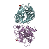
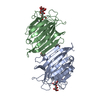
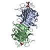
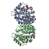
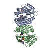
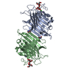
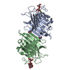
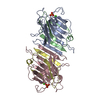
| Title | The Oligomeric Structure of Human Granzyme A Reveals the Molecular Determinants of Substrate Specificity | ||||||
 Components Components | Granzyme A | ||||||
 Keywords Keywords | HYDROLASE/HYDROLASE INHIBITOR / HYDROLASE-HYDROLASE INHIBITOR complex | ||||||
| Function / homology |  Function and homology informationgranzyme A / negative regulation of endodeoxyribonuclease activity / cytotoxic T cell pyroptotic cell death / granzyme-mediated programmed cell death signaling pathway / negative regulation of oxidoreductase activity / pyroptotic inflammatory response / negative regulation of DNA binding / protein maturation / immunological synapse / proteolysis involved in protein catabolic process ...granzyme A / negative regulation of endodeoxyribonuclease activity / cytotoxic T cell pyroptotic cell death / granzyme-mediated programmed cell death signaling pathway / negative regulation of oxidoreductase activity / pyroptotic inflammatory response / negative regulation of DNA binding / protein maturation / immunological synapse / proteolysis involved in protein catabolic process / response to bacterium / killing of cells of another organism / immune response / positive regulation of apoptotic process / serine-type endopeptidase activity / apoptotic process / protein homodimerization activity / extracellular space / nucleus / cytoplasm Function and homology informationgranzyme A / negative regulation of endodeoxyribonuclease activity / cytotoxic T cell pyroptotic cell death / granzyme-mediated programmed cell death signaling pathway / negative regulation of oxidoreductase activity / pyroptotic inflammatory response / negative regulation of DNA binding / protein maturation / immunological synapse / proteolysis involved in protein catabolic process ...granzyme A / negative regulation of endodeoxyribonuclease activity / cytotoxic T cell pyroptotic cell death / granzyme-mediated programmed cell death signaling pathway / negative regulation of oxidoreductase activity / pyroptotic inflammatory response / negative regulation of DNA binding / protein maturation / immunological synapse / proteolysis involved in protein catabolic process / response to bacterium / killing of cells of another organism / immune response / positive regulation of apoptotic process / serine-type endopeptidase activity / apoptotic process / protein homodimerization activity / extracellular space / nucleus / cytoplasmSimilarity search - Function | ||||||
| Biological species |  Homo sapiens (human) Homo sapiens (human) | ||||||
| Method | X-RAY DIFFRACTION / SYNCHROTRON / MOLECULAR REPLACEMENT / Resolution: 2.4 Å | ||||||
 Authors Authors | Bell, J.K. / Goetz, D.H. / Mahrus, S. / Harris, J.L. / Fletterick, R.J. / Craik, C.S. | ||||||
 Citation Citation | Journal: Nat.Struct.Biol. / Year: 2003 Title: The oligomeric structure of human granzyme A is a determinant of its extended substrate specificity. Authors: Bell, J.K. / Goetz, D.H. / Mahrus, S. / Harris, J.L. / Fletterick, R.J. / Craik, C.S. | ||||||
| History |
|
- Structure visualization
| Structure viewer | Molecule: MolmilJmol/JSmol |
|---|
- Downloads & links
-Download
| PDBx/mmCIF format | 1orf.cif.gz | 63.1 KB | Display | PDBx/mmCIF format |
|---|---|---|---|---|
| PDB format | pdb1orf.ent.gz | 45 KB | Display | PDB format |
| PDBx/mmJSON format | 1orf.json.gz | Tree view | PDBx/mmJSON format | |
| Others |  Other downloads Other downloads |
-Validation report
| Arichive directory | https://data.pdbj.org/pub/pdb/validation_reports/or/1orfftp://data.pdbj.org/pub/pdb/validation_reports/or/1orf | HTTPS FTP |
|---|
-Related structure data
| Related structure data |  1dstS S: Starting model for refinement |
|---|---|
| Similar structure data |


-Links
 PDBj
PDBj

- Assembly
| Deposited unit | 
| ||||||||
|---|---|---|---|---|---|---|---|---|---|
| 1 | 
| ||||||||
| Unit cell |
| ||||||||
| Details | The biological assembly is a dimer generated by the two fold axis: -x, y, 1/2-z |
-Components
| #1: Protein | / Cytotoxic T-lymphocyte proteinase 1 / Hanukkah factor / H factor / HF / Granzyme 1 / CTL tryptase / ...Cytotoxic T-lymphocyte proteinase 1 / Hanukkah factor / H factor / HF / Granzyme 1 / CTL tryptase / Fragmentin 1 Mass: 25870.066 Da / Num. of mol.: 1 / Mutation: N170E Source method: isolated from a genetically manipulated source Source: (gene. exp.) Homo sapiens (human) / Gene: GZMA / Plasmid: pPicZalphaA / Production host:  Pichia pastoris (fungus) / Strain (production host): X33 / References: UniProt: P12544, granzyme A Pichia pastoris (fungus) / Strain (production host): X33 / References: UniProt: P12544, granzyme A |
|---|---|
| #2: Chemical | ChemComp-0G6 /   Type: peptide-like, Peptide-like / Class: Inhibitor / Mass: 453.986 Da / Num. of mol.: 1 / Source method: obtained synthetically / Formula: C21H34ClN6O3 / Details: solid phase peptide synthesis / References: D-Phe-Pro-Arg-CH2Cl Type: peptide-like, Peptide-like / Class: Inhibitor / Mass: 453.986 Da / Num. of mol.: 1 / Source method: obtained synthetically / Formula: C21H34ClN6O3 / Details: solid phase peptide synthesis / References: D-Phe-Pro-Arg-CH2Cl |
| #3: Chemical | ChemComp-SO4 / Sulfate  Mass: 96.063 Da / Num. of mol.: 1 / Source method: obtained synthetically / Formula: SO4 Mass: 96.063 Da / Num. of mol.: 1 / Source method: obtained synthetically / Formula: SO4 |
| #4: Water | ChemComp-HOH / Water Mass: 18.015 Da / Num. of mol.: 128 / Source method: isolated from a natural source / Formula: H2O Mass: 18.015 Da / Num. of mol.: 128 / Source method: isolated from a natural source / Formula: H2O |
| Nonpolymer details | THE INHIBITOR IS COVALENTLY CONNECTED TO ACTIVE_SITE RESIDUES: 1) VIA A HEMIKETAL GROUP TO OG SER A ...THE INHIBITOR IS COVALENTLY |
-Experimental details
-Experiment
| Experiment | Method: X-RAY DIFFRACTION / Number of used crystals: 1 |
|---|
- Sample preparation
| Crystal | Density Matthews: 3.14 Å3/Da / Density % sol: 60.78 % | ||||||||||||||||||||||||||||||||||||||||||||||||||||||||
|---|---|---|---|---|---|---|---|---|---|---|---|---|---|---|---|---|---|---|---|---|---|---|---|---|---|---|---|---|---|---|---|---|---|---|---|---|---|---|---|---|---|---|---|---|---|---|---|---|---|---|---|---|---|---|---|---|---|
| Crystal grow | Temperature: 298 K / Method: vapor diffusion, hanging drop / pH: 8.5 Details: Tris pH 8.5, PEG 4000, lithium sulfate, VAPOR DIFFUSION, HANGING DROP, temperature 298K | ||||||||||||||||||||||||||||||||||||||||||||||||||||||||
| Crystal grow | *PLUS Temperature: 17 ℃ / pH: 6 / Method: vapor diffusion, hanging drop | ||||||||||||||||||||||||||||||||||||||||||||||||||||||||
| Components of the solutions | *PLUS
|
-Data collection
| Diffraction | Mean temperature: 100 K |
|---|---|
| Diffraction source | Source: SYNCHROTRON / Site: ALS  / Beamline: 5.0.3 / Wavelength: 1 Å / Beamline: 5.0.3 / Wavelength: 1 Å |
| Detector | Type: ADSC QUANTUM 4 / Detector: CCD / Date: Jun 2, 2002 |
| Radiation | Protocol: SINGLE WAVELENGTH / Monochromatic (M) / Laue (L): M / Scattering type: x-ray |
| Radiation wavelength | Wavelength: 1 Å / Relative weight: 1 |
| Reflection | Resolution: 2.4→20 Å / Num. all: 13319 / Num. obs: 12664 / % possible obs: 99.5 % / Observed criterion σ(F): 0 / Observed criterion σ(I): 0 |
| Reflection shell | Resolution: 2.4→2.49 Å / % possible all: 94.3 |
| Reflection | *PLUS Num. obs: 13373 / % possible obs: 99 % / Num. measured all: 175040 / Rmerge(I) obs: 0.103 |
| Reflection shell | *PLUS % possible obs: 94 % / Rmerge(I) obs: 0.417 / Mean I/σ(I) obs: 2.3 |
- Processing
| Software |
| ||||||||||||||||||||||||||||||||||||||||||||||||||||||||||||||||||||||
|---|---|---|---|---|---|---|---|---|---|---|---|---|---|---|---|---|---|---|---|---|---|---|---|---|---|---|---|---|---|---|---|---|---|---|---|---|---|---|---|---|---|---|---|---|---|---|---|---|---|---|---|---|---|---|---|---|---|---|---|---|---|---|---|---|---|---|---|---|---|---|---|
| Refinement | Method to determine structure: MOLECULAR REPLACEMENT Starting model: 1DST Resolution: 2.4→20 Å / Cor.coef. Fo:Fc: 0.943 / Cor.coef. Fo:Fc free: 0.922 / SU B: 6.903 / SU ML: 0.158 / Cross valid method: THROUGHOUT / σ(F): 0 / ESU R: 0.295 / ESU R Free: 0.223 / Stereochemistry target values: MAXIMUM LIKELIHOOD
| ||||||||||||||||||||||||||||||||||||||||||||||||||||||||||||||||||||||
| Solvent computation | Ion probe radii: 0.8 Å / Shrinkage radii: 0.8 Å / VDW probe radii: 1.4 Å / Solvent model: BABINET MODEL WITH MASK | ||||||||||||||||||||||||||||||||||||||||||||||||||||||||||||||||||||||
| Displacement parameters | Biso mean: 35.985 Å2
| ||||||||||||||||||||||||||||||||||||||||||||||||||||||||||||||||||||||
| Refine analyze |
| ||||||||||||||||||||||||||||||||||||||||||||||||||||||||||||||||||||||
| Refinement step | Cycle: LAST / Resolution: 2.4→20 Å
| ||||||||||||||||||||||||||||||||||||||||||||||||||||||||||||||||||||||
| Refine LS restraints |
| ||||||||||||||||||||||||||||||||||||||||||||||||||||||||||||||||||||||
| LS refinement shell | Resolution: 2.4→2.461 Å / Total num. of bins used: 20 /
| ||||||||||||||||||||||||||||||||||||||||||||||||||||||||||||||||||||||
| Refinement | *PLUS Highest resolution: 2.4 Å / % reflection Rfree: 5 % / Rfactor Rfree: 0.232 / Rfactor Rwork: 0.191 | ||||||||||||||||||||||||||||||||||||||||||||||||||||||||||||||||||||||
| Solvent computation | *PLUS | ||||||||||||||||||||||||||||||||||||||||||||||||||||||||||||||||||||||
| Displacement parameters | *PLUS | ||||||||||||||||||||||||||||||||||||||||||||||||||||||||||||||||||||||
| Refine LS restraints | *PLUS
|
