- PDB-1op8: Crystal Structure of Human Granzyme A -
+
Open data
ID or keywords:
Loading...
-
Basic information
Entry
Database: PDB / ID: 1op8
Title
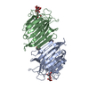
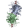
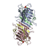
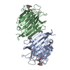
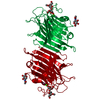
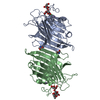
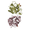
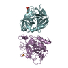
Crystal Structure of Human Granzyme A
Components
Granzyme A
Keywords
HYDROLASE / Granzyme A / Serine proteinase / Apoptosis
Function / homology
Function and homology information
granzyme A / negative regulation of endodeoxyribonuclease activity / cytotoxic T cell pyroptotic cell death / granzyme-mediated programmed cell death signaling pathway / negative regulation of oxidoreductase activity / pyroptotic inflammatory response / negative regulation of DNA binding / protein maturation / immunological synapse / proteolysis involved in protein catabolic process ...granzyme A / negative regulation of endodeoxyribonuclease activity / cytotoxic T cell pyroptotic cell death / granzyme-mediated programmed cell death signaling pathway / negative regulation of oxidoreductase activity / pyroptotic inflammatory response / negative regulation of DNA binding / protein maturation / immunological synapse / proteolysis involved in protein catabolic process / response to bacterium / killing of cells of another organism / immune response / positive regulation of apoptotic process / serine-type endopeptidase activity / apoptotic process / protein homodimerization activity / extracellular space / nucleus / cytoplasm Similarity search - Function
Serine proteases, trypsin family, histidine active site / Serine proteases, trypsin family, serine active site / Peptidase S1A, chymotrypsin family / Serine proteases, trypsin family, histidine active site. / Serine proteases, trypsin domain profile. / Serine proteases, trypsin family, serine active site. / Trypsin-like serine protease / Serine proteases, trypsin domain / Trypsin / Trypsin-like serine proteases ...Serine proteases, trypsin family, histidine active site / Serine proteases, trypsin family, serine active site / Peptidase S1A, chymotrypsin family / Serine proteases, trypsin family, histidine active site. / Serine proteases, trypsin domain profile. / Serine proteases, trypsin family, serine active site. / Trypsin-like serine protease / Serine proteases, trypsin domain / Trypsin / Trypsin-like serine proteases / Thrombin, subunit H / Peptidase S1, PA clan, chymotrypsin-like fold / Peptidase S1, PA clan / Beta Barrel / Mainly Beta Similarity search - Domain/homology
Mass: 25855.055 Da / Num. of mol.: 6 Source method: isolated from a genetically manipulated source Source: (gene. exp.) Homo sapiens (human) / Production host: Escherichia coli (E. coli) / References: UniProt: P12544, granzyme A
In the structure databanks used in Yorodumi, some data are registered as the other names, "COVID-19 virus" and "2019-nCoV". Here are the details of the virus and the list of structure data.
Jan 31, 2019. EMDB accession codes are about to change! (news from PDBe EMDB page)
EMDB accession codes are about to change! (news from PDBe EMDB page)
The allocation of 4 digits for EMDB accession codes will soon come to an end. Whilst these codes will remain in use, new EMDB accession codes will include an additional digit and will expand incrementally as the available range of codes is exhausted. The current 4-digit format prefixed with “EMD-” (i.e. EMD-XXXX) will advance to a 5-digit format (i.e. EMD-XXXXX), and so on. It is currently estimated that the 4-digit codes will be depleted around Spring 2019, at which point the 5-digit format will come into force.
The EM Navigator/Yorodumi systems omit the EMD- prefix.
Related info.:Q: What is EMD? / ID/Accession-code notation in Yorodumi/EM Navigator
Yorodumi is a browser for structure data from EMDB, PDB, SASBDB, etc.
This page is also the successor to EM Navigator detail page, and also detail information page/front-end page for Omokage search.
The word "yorodu" (or yorozu) is an old Japanese word meaning "ten thousand". "mi" (miru) is to see.
Related info.:EMDB / PDB / SASBDB / Comparison of 3 databanks / Yorodumi Search / Aug 31, 2016. New EM Navigator & Yorodumi / Yorodumi Papers / Jmol/JSmol / Function and homology information / Changes in new EM Navigator and Yorodumi
 Movie
Movie Controller


 Open data
 Basic information
 Components
 Keywords
 Function and homology information
 Authors
 Citation
 Structure visualization
 Downloads & links
 Other downloads


 PDBj
PDBj Assembly


 Mass: 96.063 Da / Num. of mol.: 12 / Source method: obtained synthetically / Formula: SO4
 Mass: 18.015 Da / Num. of mol.: 705 / Source method: isolated from a natural source / Formula: H2O
 Sample preparation
 / Beamline: BW6 / Wavelength: 1.05 Å
 Processing