 Movie
Movie Controller

[English] 日本語
 Yorodumi
- PDB-1opd: HISTIDINE-CONTAINING PROTEIN (HPR), MUTANT WITH SER 46 REPLACED B... -
+ Open data

- Basic information
| Entry | Database: PDB / ID: 1opd | ||||||
|---|---|---|---|---|---|---|---|
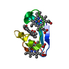
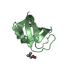
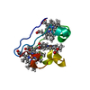
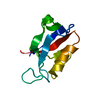
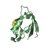
| Title | HISTIDINE-CONTAINING PROTEIN (HPR), MUTANT WITH SER 46 REPLACED BY ASP (S46D) | ||||||
 Components Components | HISTIDINE-CONTAINING PROTEIN | ||||||
 Keywords Keywords |  PHOSPHOTRANSFERASE / HISTIDINE PHOSPHOTRANSFERASE / HISTIDINE | ||||||
| Function / homology |  Function and homology information Function and homology informationphosphotransferase activity, nitrogenous group as acceptor / regulation of carbon utilization / antisigma factor binding / positive regulation of glycogen catabolic process / phosphoenolpyruvate-dependent sugar phosphotransferase system / enzyme inhibitor activity / enzyme activator activity / enzyme regulator activity / cytosolSimilarity search - Function | ||||||
| Biological species |  Escherichia coli (E. coli) Escherichia coli (E. coli) | ||||||
| Method | X-RAY DIFFRACTION / SYNCHROTRON / Resolution: 1.5 Å | ||||||
 Authors Authors | Napper, S. / Delbaere, L. / Waygood, B. | ||||||
 Citation Citation | Journal: Biochemistry / Year: 1996 Title: Mutation of serine-46 to aspartate in the histidine-containing protein of Escherichia coli mimics the inactivation by phosphorylation of serine-46 in HPrs from gram-positive bacteria. Authors: Napper, S. / Anderson, J.W. / Georges, F. / Quail, J.W. / Delbaere, L.T. / Waygood, E.B. #1: Journal: Biochemistry / Year: 1996Title: Influence of N-CAP Mutations on the Structure and Stability of Escherichia Coli Hpr Authors: Thapar, R. / Nicholson, E.M. / Rajagopal, P. / Waygood, E.B. / Scholtz, J.M. / Klevit, R.E. | ||||||
| History |
|
- Structure visualization
| Structure viewer | Molecule: MolmilJmol/JSmol |
|---|
- Downloads & links
-Download
| PDBx/mmCIF format | 1opd.cif.gz | 28.3 KB | Display | PDBx/mmCIF format |
|---|---|---|---|---|
| PDB format | pdb1opd.ent.gz | 18.4 KB | Display | PDB format |
| PDBx/mmJSON format | 1opd.json.gz | Tree view | PDBx/mmJSON format | |
| Others |  Other downloads Other downloads |
-Validation report
| Arichive directory | https://data.pdbj.org/pub/pdb/validation_reports/op/1opdftp://data.pdbj.org/pub/pdb/validation_reports/op/1opd | HTTPS FTP |
|---|
-Related structure data
| Similar structure data |
|---|

-Links
 PDBj
PDBj- Assembly
| Deposited unit | 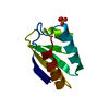
| ||||||||
|---|---|---|---|---|---|---|---|---|---|
| 1 |
| ||||||||
| Unit cell |
|
-Components
| #1: Protein | Mass: 9158.327 Da / Num. of mol.: 1 / Mutation: S46D Source method: isolated from a genetically manipulated source Source: (gene. exp.) Escherichia coli (E. coli) / Strain: ESK108 / Plasmid: PUC19 / Gene (production host): PTSH / Production host: Escherichia coli (E. coli) / Strain (production host): ESK108 / References: UniProt: P0AA04 |
|---|---|
| #2: Chemical | ChemComp-SO4 / Sulfate  Mass: 96.063 Da / Num. of mol.: 1 / Source method: obtained synthetically / Formula: SO4 Mass: 96.063 Da / Num. of mol.: 1 / Source method: obtained synthetically / Formula: SO4 |
| #3: Water | ChemComp-HOH / Water Mass: 18.015 Da / Num. of mol.: 54 / Source method: isolated from a natural source / Formula: H2O Mass: 18.015 Da / Num. of mol.: 54 / Source method: isolated from a natural source / Formula: H2O |
-Experimental details
-Experiment
| Experiment | Method: X-RAY DIFFRACTION |
|---|
- Sample preparation
| Crystal | Density Matthews: 1.82 Å3/Da / Density % sol: 32.57 % | |||||||||||||||
|---|---|---|---|---|---|---|---|---|---|---|---|---|---|---|---|---|
| Crystal grow | *PLUS Temperature: 14 ℃ / pH: 4.6 / Method: vapor diffusion, hanging drop | |||||||||||||||
| Components of the solutions | *PLUS
|
-Data collection
| Diffraction source | Source: SYNCHROTRON / Site: EMBL/DESY, HAMBURG  / Beamline: X11 / Wavelength: 0.927 / Beamline: X11 / Wavelength: 0.927 |
|---|---|
| Detector | Type: MARRESEARCH / Detector: IMAGE PLATE / Date: Jun 21, 1994 |
| Radiation | Monochromatic (M) / Laue (L): M / Scattering type: x-ray |
| Radiation wavelength | Wavelength: 0.927 Å / Relative weight: 1 |
| Reflection | Num. obs: 10525 / % possible obs: 93.4 % / Observed criterion σ(I): 0 / Redundancy: 3.2 % / Rmerge(I) obs: 0.029 |
| Reflection | *PLUS Highest resolution: 1.5 Å |
- Processing
| Software |
| ||||||||||||||||||||||||||||||||||||||||||||||||||||||||||||
|---|---|---|---|---|---|---|---|---|---|---|---|---|---|---|---|---|---|---|---|---|---|---|---|---|---|---|---|---|---|---|---|---|---|---|---|---|---|---|---|---|---|---|---|---|---|---|---|---|---|---|---|---|---|---|---|---|---|---|---|---|---|
| Refinement | Resolution: 1.5→8 Å / σ(F): 0
| ||||||||||||||||||||||||||||||||||||||||||||||||||||||||||||
| Refinement step | Cycle: LAST / Resolution: 1.5→8 Å
| ||||||||||||||||||||||||||||||||||||||||||||||||||||||||||||
| Refine LS restraints |
| ||||||||||||||||||||||||||||||||||||||||||||||||||||||||||||
| Xplor file |
| ||||||||||||||||||||||||||||||||||||||||||||||||||||||||||||
| Software | *PLUS Name: X-PLOR / Classification: refinement | ||||||||||||||||||||||||||||||||||||||||||||||||||||||||||||
| Refinement | *PLUS | ||||||||||||||||||||||||||||||||||||||||||||||||||||||||||||
| Solvent computation | *PLUS | ||||||||||||||||||||||||||||||||||||||||||||||||||||||||||||
| Displacement parameters | *PLUS |
