 Movie
Movie Controller


+ Open data

- Basic information
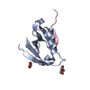
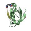
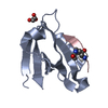
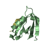
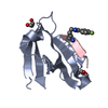
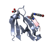
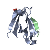
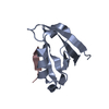
| Entry | Database: PDB / ID: 5ic3 | ||||||
|---|---|---|---|---|---|---|---|
| Title | CAL PDZ domain with peptide and inhibitor | ||||||
 Components Components |
| ||||||
 Keywords Keywords | PEPTIDE BINDING PROTEIN/INHIBITOR /  inhibitor / complex / CAL PDZ / PEPTIDE BINDING PROTEIN / PEPTIDE BINDING PROTEIN-INHIBITOR complex inhibitor / complex / CAL PDZ / PEPTIDE BINDING PROTEIN / PEPTIDE BINDING PROTEIN-INHIBITOR complex | ||||||
| Function / homology |  Function and homology information Function and homology informationnegative regulation of anion channel activity / RHO GTPases regulate CFTR trafficking / negative regulation of protein localization to cell surface / Golgi-associated vesicle membrane / Golgi to plasma membrane transport / molecular sequestering activity / trans-Golgi network transport vesicle / apical protein localization / RHOQ GTPase cycle / endoplasmic reticulum to Golgi vesicle-mediated transport ...negative regulation of anion channel activity / RHO GTPases regulate CFTR trafficking / negative regulation of protein localization to cell surface / Golgi-associated vesicle membrane / Golgi to plasma membrane transport / molecular sequestering activity / trans-Golgi network transport vesicle / apical protein localization / RHOQ GTPase cycle / endoplasmic reticulum to Golgi vesicle-mediated transport / PDZ domain binding / protein transport / symbiont-mediated suppression of host cytoplasmic pattern recognition receptor signaling pathway via inhibition of IRF3 activity / symbiont-mediated perturbation of host ubiquitin-like protein modification / transmembrane transporter binding / host cell cytoplasm / postsynaptic density / symbiont-mediated suppression of host type I interferon-mediated signaling pathway / lysosomal membrane / virus-mediated perturbation of host defense response / Golgi membrane / negative regulation of DNA-templated transcription / DNA-templated transcription / dendrite / host cell nucleus / Golgi apparatus / protein-containing complex / DNA binding / zinc ion binding / membrane / identical protein binding / plasma membrane / cytoplasmSimilarity search - Function | ||||||
| Biological species |  Homo sapiens (human) Homo sapiens (human) Alphapapillomavirus 7 Alphapapillomavirus 7 | ||||||
| Method | X-RAY DIFFRACTION / SYNCHROTRON / MOLECULAR REPLACEMENT / Resolution: 1.7 Å | ||||||
 Authors Authors | Zhao, Y. / Madden, D.R. | ||||||
| Funding support |  United States, 1items United States, 1items
| ||||||
 Citation Citation | Journal: To Be Published Title: CAL PDZ domain with peptide and inhibitor Authors: Zhao, Y. / Madden, D.R. | ||||||
| History |
|
- Structure visualization
| Structure viewer | Molecule: MolmilJmol/JSmol |
|---|
- Downloads & links
-Download
| PDBx/mmCIF format | 5ic3.cif.gz | 86 KB | Display | PDBx/mmCIF format |
|---|---|---|---|---|
| PDB format | pdb5ic3.ent.gz | 65.4 KB | Display | PDB format |
| PDBx/mmJSON format | 5ic3.json.gz | Tree view | PDBx/mmJSON format | |
| Others |  Other downloads Other downloads |
-Validation report
| Arichive directory | https://data.pdbj.org/pub/pdb/validation_reports/ic/5ic3ftp://data.pdbj.org/pub/pdb/validation_reports/ic/5ic3 | HTTPS FTP |
|---|
-Related structure data
| Related structure data |  4e34S S: Starting model for refinement |
|---|---|
| Similar structure data |


-Links
 PDBj
PDBj

- Assembly
| Deposited unit | 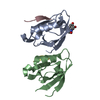
| ||||||||
|---|---|---|---|---|---|---|---|---|---|
| 1 | 
| ||||||||
| 2 | 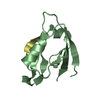
| ||||||||
| Unit cell |
|
-Components
| #1: Protein | Mass: 9353.722 Da / Num. of mol.: 2 / Fragment: UNP residues 284-370 Source method: isolated from a genetically manipulated source Source: (gene. exp.) Homo sapiens (human) / Gene: GOPC, CAL, FIG / Plasmid: pET16 / Production host:  Escherichia coli (E. coli) / Strain (production host): BL21 / Variant (production host): DE3 RIL / References: UniProt: Q9HD26 Escherichia coli (E. coli) / Strain (production host): BL21 / Variant (production host): DE3 RIL / References: UniProt: Q9HD26#2: Protein/peptide | Mass: 1345.554 Da / Num. of mol.: 2 / Source method: obtained synthetically / Source: (synth.) Alphapapillomavirus 7 / References: UniProt: P06463*PLUS#3: Chemical | ChemComp-PQR / |   Mass: 168.150 Da / Num. of mol.: 1 Mass: 168.150 Da / Num. of mol.: 1Source method: isolated from a genetically manipulated source Formula: C7H8N2O3 #4: Water | ChemComp-HOH / | Water Mass: 18.015 Da / Num. of mol.: 144 / Source method: isolated from a natural source / Formula: H2O Mass: 18.015 Da / Num. of mol.: 144 / Source method: isolated from a natural source / Formula: H2OSequence details | the numbering of the sequence is made according to the isofrom 2 of CAL | |
|---|
-Experimental details
-Experiment
| Experiment | Method: X-RAY DIFFRACTION / Number of used crystals: 1 |
|---|
- Sample preparation
| Crystal | Density Matthews: 1.87 Å3/Da / Density % sol: 34.33 % |
|---|---|
| Crystal grow | Temperature: 291 K / Method: vapor diffusion, hanging drop / pH: 7.4 / Details: 35% PEG3350, 100mM NaCl, 100mM Tris pH7.4 |
-Data collection
| Diffraction | Mean temperature: 100 K |
|---|---|
| Diffraction source | Source: SYNCHROTRON / Site: NSLS / Beamline: X6A / Wavelength: 0.9795 Å |
| Detector | Type: ADSC QUANTUM 270 / Detector: CCD / Date: Feb 24, 2013 |
| Radiation | Monochromator: Si(111) / Protocol: SINGLE WAVELENGTH / Monochromatic (M) / Laue (L): M / Scattering type: x-ray |
| Radiation wavelength | Wavelength: 0.9795 Å / Relative weight: 1 |
| Reflection | Resolution: 1.7→19.29 Å / Num. obs: 17472 / % possible obs: 99.9 % / Redundancy: 7.51 % / Rmerge(I) obs: 0.062 / Net I/σ(I): 23.78 |
| Reflection shell | Highest resolution: 1.7 Å / Rmerge(I) obs: 0.686 |
- Processing
| Software |
| |||||||||||||||||||||||||||||||||||||||||||||||||
|---|---|---|---|---|---|---|---|---|---|---|---|---|---|---|---|---|---|---|---|---|---|---|---|---|---|---|---|---|---|---|---|---|---|---|---|---|---|---|---|---|---|---|---|---|---|---|---|---|---|---|
| Refinement | Method to determine structure: MOLECULAR REPLACEMENT / Starting model: 4.0E+34 / Resolution: 1.7→19.29 Å / SU ML: 0.24 / Cross valid method: FREE R-VALUE / σ(F): 2 / Phase error: 22.49
| |||||||||||||||||||||||||||||||||||||||||||||||||
| Solvent computation | Shrinkage radii: 0.9 Å / VDW probe radii: 1.11 Å / Bsol: 52.15 Å2 / ksol: 0.42 e/Å3 | |||||||||||||||||||||||||||||||||||||||||||||||||
| Displacement parameters |
| |||||||||||||||||||||||||||||||||||||||||||||||||
| Refinement step | Cycle: LAST / Resolution: 1.7→19.29 Å
| |||||||||||||||||||||||||||||||||||||||||||||||||
| Refine LS restraints |
| |||||||||||||||||||||||||||||||||||||||||||||||||
| LS refinement shell |
|
