 Movie
Movie Controller

[English] 日本語
 Yorodumi
- PDB-1olo: Hexameric Replicative DNA Helicase RepA from Plasmid RSF1010 - Cu... -
+ Open data

- Basic information
| Entry | Database: PDB / ID: 1olo | ||||||
|---|---|---|---|---|---|---|---|
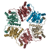
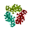
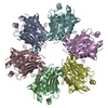
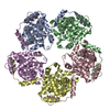
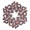
| Title | Hexameric Replicative DNA Helicase RepA from Plasmid RSF1010 - Cubic Crystal Structure | ||||||
 Components Components | REGULATORY PROTEIN REPA | ||||||
 Keywords Keywords |  TRANSCRIPTION / DNA HELICASE / ATPASE / MOTOR PROTEIN TRANSCRIPTION / DNA HELICASE / ATPASE / MOTOR PROTEIN | ||||||
| Function / homology |  Function and homology information Function and homology information | ||||||
| Biological species |  ESCHERICHIA COLI (E. coli) ESCHERICHIA COLI (E. coli) | ||||||
| Method | X-RAY DIFFRACTION / MAD / Resolution: 2.55 Å | ||||||
 Authors Authors | Niedenzu, T. / Saenger, W. | ||||||
 Citation Citation | Journal: Nucleic Acids Res. / Year: 2003 Title: Hexameric Rsf1010 Helicase Repa: The Structural and Functional Importance of Single Amino Acid Residues Authors: Ziegelin, G. / Niedenzu, T. / Lurz, R. / Saenger, W. / Lanka, E. | ||||||
| History |
|
- Structure visualization
| Structure viewer | Molecule: MolmilJmol/JSmol |
|---|
- Downloads & links
-Download
| PDBx/mmCIF format | 1olo.cif.gz | 116.6 KB | Display | PDBx/mmCIF format |
|---|---|---|---|---|
| PDB format | pdb1olo.ent.gz | 91.8 KB | Display | PDB format |
| PDBx/mmJSON format | 1olo.json.gz | Tree view | PDBx/mmJSON format | |
| Others |  Other downloads Other downloads |
-Validation report
| Arichive directory | https://data.pdbj.org/pub/pdb/validation_reports/ol/1oloftp://data.pdbj.org/pub/pdb/validation_reports/ol/1olo | HTTPS FTP |
|---|
-Related structure data
| Related structure data | 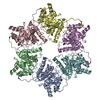 1g8yS S: Starting model for refinement |
|---|---|
| Similar structure data |


-Links
 PDBj
PDBj
- Assembly
| Deposited unit | 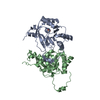
| ||||||||
|---|---|---|---|---|---|---|---|---|---|
| 1 | 
| ||||||||
| Unit cell |
| ||||||||
| Components on special symmetry positions |
| ||||||||
| Details | THE ASSEMBLY DESCRIBED BELOW IS A HOMOHEXAMERIC ASSEMBLY. |
-Components
| #1: Protein | Mass: 29945.189 Da / Num. of mol.: 2 Source method: isolated from a genetically manipulated source Source: (gene. exp.) ESCHERICHIA COLI (E. coli) / Description: BROAD HOST RANGE PLASMID RSF1010 / Plasmid: PTACREP-A / Production host: ESCHERICHIA COLI (E. coli) / Strain (production host): XL1-BLUE / References: UniProt: P20356#2: Chemical | Sulfate  Mass: 96.063 Da / Num. of mol.: 2 / Source method: obtained synthetically / Formula: SO4 Mass: 96.063 Da / Num. of mol.: 2 / Source method: obtained synthetically / Formula: SO4#3: Water | ChemComp-HOH / | Water Mass: 18.015 Da / Num. of mol.: 421 / Source method: isolated from a natural source / Formula: H2O Mass: 18.015 Da / Num. of mol.: 421 / Source method: isolated from a natural source / Formula: H2O |
|---|
-Experimental details
-Experiment
| Experiment | Method: X-RAY DIFFRACTION / Number of used crystals: 1 |
|---|
- Sample preparation
| Crystal | Density Matthews: 4.8 Å3/Da / Density % sol: 74.4 % | ||||||||||||||||||||||||||||||||||||||||||||||||||||||||
|---|---|---|---|---|---|---|---|---|---|---|---|---|---|---|---|---|---|---|---|---|---|---|---|---|---|---|---|---|---|---|---|---|---|---|---|---|---|---|---|---|---|---|---|---|---|---|---|---|---|---|---|---|---|---|---|---|---|
| Crystal grow | Temperature: 291 K / Method: vapor diffusion, sitting drop / pH: 7.5 Details: SITTING DROP VAPOR DIFFUSION AT 291K. PROTEIN STOCK: 26MG/ML REPA, 10MM TRIS-HCL PH8.0, 150MM NACL, 0.1MM EDTA. PRECIPITANT: 100MM TRIS-HCL PH 7.5, 26% PEG400, 100MM MGSO4 | ||||||||||||||||||||||||||||||||||||||||||||||||||||||||
| Crystal grow | *PLUS pH: 8 / Method: vapor diffusion, sitting drop | ||||||||||||||||||||||||||||||||||||||||||||||||||||||||
| Components of the solutions | *PLUS
|
-Data collection
| Diffraction | Mean temperature: 100 K |
|---|---|
| Diffraction source | Source: ROTATING ANODE / Type: ENRAF-NONIUS FR571 / Wavelength: 1.5418 |
| Detector | Type: MARRESEARCH / Detector: IMAGE PLATE / Date: Dec 15, 2000 / Details: MIRRORS |
| Radiation | Monochromator: GRAPHITE / Protocol: MAD / Monochromatic (M) / Laue (L): M / Scattering type: x-ray |
| Radiation wavelength | Wavelength: 1.5418 Å / Relative weight: 1 |
| Reflection | Resolution: 2.52→150 Å / Num. obs: 40046 / % possible obs: 99.1 % / Redundancy: 7.64 % / Rmerge(I) obs: 0.106 / Net I/σ(I): 13.6 |
| Reflection shell | Resolution: 2.52→2.58 Å / Redundancy: 4.1 % / Rmerge(I) obs: 0.6 / Mean I/σ(I) obs: 3.3 / % possible all: 89.7 |
| Reflection | *PLUS Highest resolution: 2.52 Å / Num. measured all: 305969 / Rmerge(I) obs: 0.106 |
| Reflection shell | *PLUS Rmerge(I) obs: 0.62 / Mean I/σ(I) obs: 3.3 |
- Processing
| Software |
| ||||||||||||||||||||||||||||||||||||||||||||||||||||||||||||||||||||||||||||||||||||||||||||||||||||||||||||||||||||||||||||||||||||||||||||||||||||||||||||||||||||||||||||||||||||||
|---|---|---|---|---|---|---|---|---|---|---|---|---|---|---|---|---|---|---|---|---|---|---|---|---|---|---|---|---|---|---|---|---|---|---|---|---|---|---|---|---|---|---|---|---|---|---|---|---|---|---|---|---|---|---|---|---|---|---|---|---|---|---|---|---|---|---|---|---|---|---|---|---|---|---|---|---|---|---|---|---|---|---|---|---|---|---|---|---|---|---|---|---|---|---|---|---|---|---|---|---|---|---|---|---|---|---|---|---|---|---|---|---|---|---|---|---|---|---|---|---|---|---|---|---|---|---|---|---|---|---|---|---|---|---|---|---|---|---|---|---|---|---|---|---|---|---|---|---|---|---|---|---|---|---|---|---|---|---|---|---|---|---|---|---|---|---|---|---|---|---|---|---|---|---|---|---|---|---|---|---|---|---|---|
| Refinement | Method to determine structure: MAD Starting model: PDB ENTRY 1G8Y Resolution: 2.55→25 Å / Cor.coef. Fo:Fc: 0.957 / Cor.coef. Fo:Fc free: 0.939 / SU B: 5.671 / SU ML: 0.123 / TLS residual ADP flag: LIKELY RESIDUAL / Cross valid method: THROUGHOUT / ESU R: 0.207 / ESU R Free: 0.183 / Stereochemistry target values: MAXIMUM LIKELIHOOD
| ||||||||||||||||||||||||||||||||||||||||||||||||||||||||||||||||||||||||||||||||||||||||||||||||||||||||||||||||||||||||||||||||||||||||||||||||||||||||||||||||||||||||||||||||||||||
| Solvent computation | Ion probe radii: 0.8 Å / Shrinkage radii: 0.8 Å / VDW probe radii: 1.4 Å / Solvent model: BABINET MODEL PLUS MASK | ||||||||||||||||||||||||||||||||||||||||||||||||||||||||||||||||||||||||||||||||||||||||||||||||||||||||||||||||||||||||||||||||||||||||||||||||||||||||||||||||||||||||||||||||||||||
| Displacement parameters | Biso mean: 34.09 Å2 | ||||||||||||||||||||||||||||||||||||||||||||||||||||||||||||||||||||||||||||||||||||||||||||||||||||||||||||||||||||||||||||||||||||||||||||||||||||||||||||||||||||||||||||||||||||||
| Refinement step | Cycle: LAST / Resolution: 2.55→25 Å
| ||||||||||||||||||||||||||||||||||||||||||||||||||||||||||||||||||||||||||||||||||||||||||||||||||||||||||||||||||||||||||||||||||||||||||||||||||||||||||||||||||||||||||||||||||||||
| Refine LS restraints |
|