 Movie
Movie Controller

[English] 日本語
 Yorodumi
- PDB-1g8y: CRYSTAL STRUCTURE OF THE HEXAMERIC REPLICATIVE HELICASE REPA OF P... -
+ Open data

- Basic information
| Entry | Database: PDB / ID: 1g8y | ||||||
|---|---|---|---|---|---|---|---|
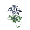
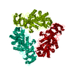
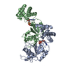
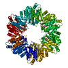
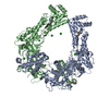
| Title | CRYSTAL STRUCTURE OF THE HEXAMERIC REPLICATIVE HELICASE REPA OF PLASMID RSF1010 | ||||||
 Components Components | REGULATORY PROTEIN REPA | ||||||
 Keywords Keywords |  TRANSCRIPTION / P-loop TRANSCRIPTION / P-loop | ||||||
| Function / homology |  Function and homology information Function and homology information | ||||||
| Biological species |  Escherichia coli (E. coli) Escherichia coli (E. coli) | ||||||
| Method | X-RAY DIFFRACTION / SYNCHROTRON / MAD / Resolution: 2.4 Å | ||||||
 Authors Authors | Niedenzu, T. / Roeleke, D. / Bains, G. / Scherzinger, E. / Saenger, W. | ||||||
 Citation Citation | Journal: J.Mol.Biol. / Year: 2001 Title: Crystal structure of the hexameric replicative helicase RepA of plasmid RSF1010. Authors: Niedenzu, T. / Roleke, D. / Bains, G. / Scherzinger, E. / Saenger, W. | ||||||
| History |
|
- Structure visualization
| Structure viewer | Molecule: MolmilJmol/JSmol |
|---|
- Downloads & links
-Download
| PDBx/mmCIF format | 1g8y.cif.gz | 547.8 KB | Display | PDBx/mmCIF format |
|---|---|---|---|---|
| PDB format | pdb1g8y.ent.gz | 453.6 KB | Display | PDB format |
| PDBx/mmJSON format | 1g8y.json.gz | Tree view | PDBx/mmJSON format | |
| Others |  Other downloads Other downloads |
-Validation report
| Arichive directory | https://data.pdbj.org/pub/pdb/validation_reports/g8/1g8yftp://data.pdbj.org/pub/pdb/validation_reports/g8/1g8y | HTTPS FTP |
|---|
-Related structure data
| Similar structure data |
|---|
-Links
 PDBj
PDBj- Assembly
| Deposited unit | 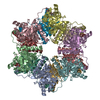
| ||||||||||
|---|---|---|---|---|---|---|---|---|---|---|---|
| 1 | 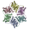
| ||||||||||
| 2 | 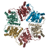
| ||||||||||
| Unit cell |
| ||||||||||
| Details | The biological assembly is one of the two hexamers in the asymmetric unit: chains a-f or g-l |
-Components
| #1: Protein | Mass: 29945.189 Da / Num. of mol.: 12 Source method: isolated from a genetically manipulated source Source: (gene. exp.) Escherichia coli (E. coli) / Gene: REPA ON PLASMID INCQ RSF1010 / Plasmid: PTACREP-A / Production host: Escherichia coli (E. coli) / Strain (production host): XL1-BLUE / References: UniProt: P20356#2: Water | ChemComp-HOH / | Water Mass: 18.015 Da / Num. of mol.: 480 / Source method: isolated from a natural source / Formula: H2O Mass: 18.015 Da / Num. of mol.: 480 / Source method: isolated from a natural source / Formula: H2O |
|---|
-Experimental details
-Experiment
| Experiment | Method: X-RAY DIFFRACTION / Number of used crystals: 1 |
|---|
- Sample preparation
| Crystal | Density Matthews: 2.9 Å3/Da / Density % sol: 57 % | ||||||||||||||||||||||||||||||||||||||||||||||||
|---|---|---|---|---|---|---|---|---|---|---|---|---|---|---|---|---|---|---|---|---|---|---|---|---|---|---|---|---|---|---|---|---|---|---|---|---|---|---|---|---|---|---|---|---|---|---|---|---|---|
| Crystal grow | Temperature: 291 K / Method: vapor diffusion, hanging drop / pH: 6 Details: PEG 5000 monomethyl ether, 2-methyl-2,4-pentanediol, NaCl, sodium citrate, pH 6.0, VAPOR DIFFUSION, HANGING DROP, temperature 291K | ||||||||||||||||||||||||||||||||||||||||||||||||
| Crystal grow | *PLUS pH: 8 | ||||||||||||||||||||||||||||||||||||||||||||||||
| Components of the solutions | *PLUS
|
-Data collection
| Diffraction | Mean temperature: 100 K |
|---|---|
| Diffraction source | Source: SYNCHROTRON / Site: EMBL/DESY, HAMBURG  / Beamline: X11 / Wavelength: 0.9059 Å / Beamline: X11 / Wavelength: 0.9059 Å |
| Detector | Type: MARRESEARCH / Detector: IMAGE PLATE / Date: Oct 22, 1998 |
| Radiation | Monochromator: bent single-crystal germanium triangular monochromator Protocol: SINGLE WAVELENGTH / Monochromatic (M) / Laue (L): M / Scattering type: x-ray |
| Radiation wavelength | Wavelength: 0.9059 Å / Relative weight: 1 |
| Reflection | Resolution: 2.4→50 Å / Num. all: 149192 / Num. obs: 149192 / % possible obs: 94.6 % / Observed criterion σ(F): 0 / Observed criterion σ(I): -3 / Redundancy: 2.53 % / Biso Wilson estimate: 26.8 Å2 / Rmerge(I) obs: 0.037 / Net I/σ(I): 18.9 |
| Reflection shell | Resolution: 2.4→2.46 Å / Redundancy: 2.46 % / Rmerge(I) obs: 0.117 / Mean I/σ(I) obs: 6.9 / Num. unique all: 8579 / % possible all: 81.7 |
| Reflection | *PLUS Num. measured all: 377676 / Rmerge(I) obs: 0.034 |
| Reflection shell | *PLUS % possible obs: 81.7 % |
- Processing
| Software |
| |||||||||||||||||||||||||
|---|---|---|---|---|---|---|---|---|---|---|---|---|---|---|---|---|---|---|---|---|---|---|---|---|---|---|
| Refinement | Method to determine structure: MAD / Resolution: 2.4→30 Å / Isotropic thermal model: RESTRAINED / Cross valid method: THROUGHOUT / σ(F): 0 / σ(I): 0 / Stereochemistry target values: Engh & Huber
| |||||||||||||||||||||||||
| Displacement parameters |
| |||||||||||||||||||||||||
| Refine analyze |
| |||||||||||||||||||||||||
| Refinement step | Cycle: LAST / Resolution: 2.4→30 Å
| |||||||||||||||||||||||||
| Refine LS restraints |
| |||||||||||||||||||||||||
| LS refinement shell | Resolution: 2.4→2.49 Å / Rfactor Rfree error: 0.02
| |||||||||||||||||||||||||
| Software | *PLUS Name: CNS / Classification: refinement | |||||||||||||||||||||||||
| Refinement | *PLUS Lowest resolution: 30 Å / σ(F): 0 / % reflection Rfree: 2.1 % / Rfactor obs: 0.221 | |||||||||||||||||||||||||
| Solvent computation | *PLUS | |||||||||||||||||||||||||
| Displacement parameters | *PLUS | |||||||||||||||||||||||||
| Refine LS restraints | *PLUS Type: c_angle_deg / Dev ideal: 1.5 | |||||||||||||||||||||||||
| LS refinement shell | *PLUS Rfactor Rfree: 0.32 / Rfactor Rwork: 0.261 |
