 Movie
Movie Controller


+ Open data

- Basic information
| Entry | Database: PDB / ID: 4j4u | ||||||
|---|---|---|---|---|---|---|---|
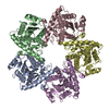
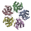
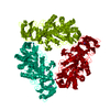
| Title | Pentamer SFTSVN | ||||||
 Components Components | Nucleocapsid protein Virus Virus | ||||||
 Keywords Keywords | VIRAL PROTEIN / nucleocapsid protein / nucleoprotein / nucleocapsid | ||||||
| Function / homology |  Function and homology information Function and homology informationhost cell endoplasmic reticulum-Golgi intermediate compartment / host cell Golgi apparatus / viral nucleocapsid / ribonucleoprotein complex / host cell nucleus / RNA bindingSimilarity search - Function | ||||||
| Biological species |  Phlebovirus Phlebovirus | ||||||
| Method | X-RAY DIFFRACTION / SYNCHROTRON / MOLECULAR REPLACEMENT / Resolution: 2.803 Å | ||||||
 Authors Authors | Jiao, L. / Ouyang, S. / Liang, M. / Niu, F. / Shaw, N. / Wu, W. / Ding, W. / Jin, C. / Zhu, Y. / Zhang, F. ...Jiao, L. / Ouyang, S. / Liang, M. / Niu, F. / Shaw, N. / Wu, W. / Ding, W. / Jin, C. / Zhu, Y. / Zhang, F. / Wang, T. / Li, C. / Zuo, X. / Luan, C.H. / Li, D. / Liu, Z.J. | ||||||
 Citation Citation | Journal: J.Virol. / Year: 2013 Title: Structure of severe Fever with thrombocytopenia syndrome virus nucleocapsid protein in complex with suramin reveals therapeutic potential Authors: Jiao, L. / Ouyang, S. / Liang, M. / Niu, F. / Shaw, N. / Wu, W. / Ding, W. / Jin, C. / Peng, Y. / Zhu, Y. / Zhang, F. / Wang, T. / Li, C. / Zuo, X. / Luan, C.H. / Li, D. / Liu, Z.J. | ||||||
| History |
|
- Structure visualization
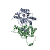
| Structure viewer | Molecule: MolmilJmol/JSmol |
|---|
- Downloads & links
-Download
| PDBx/mmCIF format | 4j4u.cif.gz | 462 KB | Display | PDBx/mmCIF format |
|---|---|---|---|---|
| PDB format | pdb4j4u.ent.gz | 381.6 KB | Display | PDB format |
| PDBx/mmJSON format | 4j4u.json.gz | Tree view | PDBx/mmJSON format | |
| Others |  Other downloads Other downloads |
-Validation report
| Arichive directory | https://data.pdbj.org/pub/pdb/validation_reports/j4/4j4uftp://data.pdbj.org/pub/pdb/validation_reports/j4/4j4u | HTTPS FTP |
|---|
-Related structure data
| Related structure data | 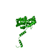 4j4rSC  4j4sC 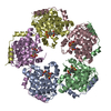 4j4vC  4j4wC  4j4xC  4j4yC S: Starting model for refinement C: citing same article ( |
|---|---|
| Similar structure data |

-Links
 PDBj
PDBj- Assembly
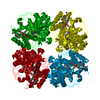
| Deposited unit | 
| ||||||||
|---|---|---|---|---|---|---|---|---|---|
| 1 |
| ||||||||
| Unit cell |
|
-Components
| #1: Protein | Virus / SFTSVN Mass: 27289.492 Da / Num. of mol.: 5 Source method: isolated from a genetically manipulated source Source: (gene. exp.) Phlebovirus / Strain: JS2010-018 / Production host:  Escherichia coli (E. coli) / References: UniProt: I6WJ72 Escherichia coli (E. coli) / References: UniProt: I6WJ72#2: Water | ChemComp-HOH / | Water Mass: 18.015 Da / Num. of mol.: 59 / Source method: isolated from a natural source / Formula: H2O Mass: 18.015 Da / Num. of mol.: 59 / Source method: isolated from a natural source / Formula: H2O |
|---|
-Experimental details
-Experiment
| Experiment | Method: X-RAY DIFFRACTION / Number of used crystals: 1 |
|---|
- Sample preparation
| Crystal | Density Matthews: 2.95 Å3/Da / Density % sol: 58.33 % |
|---|
-Data collection
| Diffraction | Mean temperature: 100 K |
|---|---|
| Diffraction source | Source: SYNCHROTRON / Site: SSRF  / Beamline: BL17U / Wavelength: 0.98 Å / Beamline: BL17U / Wavelength: 0.98 Å |
| Detector | Type: ADSC QUANTUM 315 / Detector: CCD / Date: Nov 26, 2011 |
| Radiation | Monochromator: SAGITALLY FOCUSED Si(111) / Protocol: SINGLE WAVELENGTH / Monochromatic (M) / Laue (L): M / Scattering type: x-ray |
| Radiation wavelength | Wavelength: 0.98 Å / Relative weight: 1 |
| Reflection | Resolution: 2.8→43.81 Å / Num. all: 35894 / Num. obs: 38804 / % possible obs: 92.5 % / Observed criterion σ(F): 2 / Observed criterion σ(I): 2 / Biso Wilson estimate: 42.86 Å2 |
| Reflection shell | Resolution: 2.8→2.9 Å / % possible all: 92.4 |
- Processing
| Software | Name: PHENIX / Version: (phenix.refine: 1.8.1_1168) / Classification: refinement | |||||||||||||||||||||||||||||||||||||||||||||||||||||||||||||||||||||||||||||||||||||||||||||||||||||||||
|---|---|---|---|---|---|---|---|---|---|---|---|---|---|---|---|---|---|---|---|---|---|---|---|---|---|---|---|---|---|---|---|---|---|---|---|---|---|---|---|---|---|---|---|---|---|---|---|---|---|---|---|---|---|---|---|---|---|---|---|---|---|---|---|---|---|---|---|---|---|---|---|---|---|---|---|---|---|---|---|---|---|---|---|---|---|---|---|---|---|---|---|---|---|---|---|---|---|---|---|---|---|---|---|---|---|---|
| Refinement | Method to determine structure: MOLECULAR REPLACEMENT Starting model: 4J4R Resolution: 2.803→43.808 Å / Occupancy max: 1 / Occupancy min: 1 / FOM work R set: 0.7714 / SU ML: 0.43 / σ(F): 1.36 / Phase error: 29.92 / Stereochemistry target values: ML
| |||||||||||||||||||||||||||||||||||||||||||||||||||||||||||||||||||||||||||||||||||||||||||||||||||||||||
| Solvent computation | Shrinkage radii: 0.9 Å / VDW probe radii: 1.11 Å / Solvent model: FLAT BULK SOLVENT MODEL | |||||||||||||||||||||||||||||||||||||||||||||||||||||||||||||||||||||||||||||||||||||||||||||||||||||||||
| Displacement parameters | Biso max: 167.1 Å2 / Biso mean: 36.5286 Å2 / Biso min: 7.59 Å2 | |||||||||||||||||||||||||||||||||||||||||||||||||||||||||||||||||||||||||||||||||||||||||||||||||||||||||
| Refinement step | Cycle: LAST / Resolution: 2.803→43.808 Å
| |||||||||||||||||||||||||||||||||||||||||||||||||||||||||||||||||||||||||||||||||||||||||||||||||||||||||
| Refine LS restraints |
| |||||||||||||||||||||||||||||||||||||||||||||||||||||||||||||||||||||||||||||||||||||||||||||||||||||||||
| LS refinement shell | Refine-ID: X-RAY DIFFRACTION / Total num. of bins used: 14
| |||||||||||||||||||||||||||||||||||||||||||||||||||||||||||||||||||||||||||||||||||||||||||||||||||||||||
| Refinement TLS params. | Method: refined / Origin x: -15.1412 Å / Origin y: -7.579 Å / Origin z: 22.1952 Å
| |||||||||||||||||||||||||||||||||||||||||||||||||||||||||||||||||||||||||||||||||||||||||||||||||||||||||
| Refinement TLS group | Selection details: all |
