 Movie
Movie Controller


+ Open data

- Basic information
| Entry | Database: PDB / ID: 1ode | ||||||
|---|---|---|---|---|---|---|---|
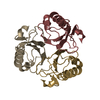
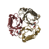
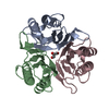
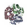
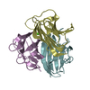
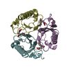
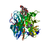
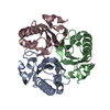
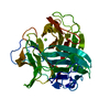
| Title | Crystal Analysis Of Chorismate Mutase From Thermus Thermophilus. | ||||||
 Components Components | CHORISMATE MUTASE | ||||||
 Keywords Keywords | ISOMERASE / CHORISMATE MUTASE / SHIKIMATE PATHWAY / MUTANT / RIKEN STRUCTURAL GENOMICS/PROTEOMICS INITIATIVE / RSGI / STRUCTURAL GENOMICS | ||||||
| Function / homology |  Function and homology information Function and homology informationchorismate metabolic process / chorismate mutase / chorismate mutase activity / aromatic amino acid family biosynthetic process / amino acid biosynthetic process / cytoplasmSimilarity search - Function | ||||||
| Biological species |   THERMUS THERMOPHILUS (bacteria) THERMUS THERMOPHILUS (bacteria) | ||||||
| Method | X-RAY DIFFRACTION / SYNCHROTRON / MOLECULAR REPLACEMENT / Resolution: 1.65 Å | ||||||
 Authors Authors | Inagaki, E. / Miyano, M. / Tahirov, T.H. | ||||||
 Citation Citation | Journal: To be Published Title: The Crystal Structure of Chorismate Mutase from Thermus Thermophilus Authors: Inagaki, E. / Kuramitsu, S. / Yokoyama, S. / Miyano, M. / Tahirov, T.H. | ||||||
| History |
|
- Structure visualization
| Structure viewer | Molecule: MolmilJmol/JSmol |
|---|
- Downloads & links
-Download
| PDBx/mmCIF format | 1ode.cif.gz | 85.3 KB | Display | PDBx/mmCIF format |
|---|---|---|---|---|
| PDB format | pdb1ode.ent.gz | 65.6 KB | Display | PDB format |
| PDBx/mmJSON format | 1ode.json.gz | Tree view | PDBx/mmJSON format | |
| Others |  Other downloads Other downloads |
-Validation report
| Arichive directory | https://data.pdbj.org/pub/pdb/validation_reports/od/1odeftp://data.pdbj.org/pub/pdb/validation_reports/od/1ode | HTTPS FTP |
|---|
-Related structure data
| Related structure data | 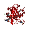 1ufyC 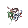 1dbfS S: Starting model for refinement C: citing same article ( |
|---|---|
| Similar structure data |

-Links
 PDBj
PDBj
- Assembly
| Deposited unit | 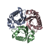
| ||||||||
|---|---|---|---|---|---|---|---|---|---|
| 1 |
| ||||||||
| Unit cell |
|
-Components
| #1: Protein | Mass: 13666.668 Da / Num. of mol.: 3 / Mutation: YES Source method: isolated from a genetically manipulated source Details: MUTATION OF PHE 55 TO SER / Source: (gene. exp.) THERMUS THERMOPHILUS (bacteria) / Strain: HB8 / Plasmid: PET11A / Production host: ESCHERICHIA COLI (E. coli) / Strain (production host): BL21(DE3) / References: UniProt: Q84FH6*PLUS, chorismate mutase#2: Water | ChemComp-HOH / | Water Mass: 18.015 Da / Num. of mol.: 332 / Source method: isolated from a natural source / Formula: H2O Mass: 18.015 Da / Num. of mol.: 332 / Source method: isolated from a natural source / Formula: H2OCompound details | ENGINEERED | Sequence details | COMPOUND ENGINEERED MUTATION IN CHAIN A, PHE 55 TO SER 55 ENGINEERED MUTATION IN CHAIN B, PHE 55 TO ...COMPOUND ENGINEERED | |
|---|
-Experimental details
-Experiment
| Experiment | Method: X-RAY DIFFRACTION / Number of used crystals: 1 |
|---|
- Sample preparation
| Crystal | Density Matthews: 1.9 Å3/Da / Density % sol: 36.9 % |
|---|---|
| Crystal grow | Method: microbatch / pH: 8.4 Details: PROTEIN WAS CRYSTALLIZED USING MICRO BATCH METHOD UNDER OIL FROM 27.5% PEG 4000, 10% DIOXANE AND 100 MM, TRIS/HCL(PH 8.4). |
-Data collection
| Diffraction | Mean temperature: 100 K |
|---|---|
| Diffraction source | Source: SYNCHROTRON / Site: SPring-8  / Beamline: BL45XU / Wavelength: 1.02 / Beamline: BL45XU / Wavelength: 1.02 |
| Detector | Type: RIGAKU IMAGE PLATE RAXIS V / Detector: IMAGE PLATE / Date: Oct 15, 2002 / Details: CYLINDRICAL BEND MIRROR |
| Radiation | Monochromator: DIAMOND / Protocol: SINGLE WAVELENGTH / Monochromatic (M) / Laue (L): M / Scattering type: x-ray |
| Radiation wavelength | Wavelength: 1.02 Å / Relative weight: 1 |
| Reflection | Resolution: 1.65→50 Å / Num. obs: 174516 / % possible obs: 99.5 % / Observed criterion σ(I): -0.5 / Redundancy: 4.4 % / Biso Wilson estimate: 15.4 Å2 / Rmerge(I) obs: 0.064 / Net I/σ(I): 23.5 |
| Reflection shell | Resolution: 1.65→1.71 Å / Rmerge(I) obs: 0.383 / Mean I/σ(I) obs: 2.4 / % possible all: 99.5 |
- Processing
| Software |
| ||||||||||||||||||||||||||||||||||||||||||||||||||||||||||||||||||||||||||||||||
|---|---|---|---|---|---|---|---|---|---|---|---|---|---|---|---|---|---|---|---|---|---|---|---|---|---|---|---|---|---|---|---|---|---|---|---|---|---|---|---|---|---|---|---|---|---|---|---|---|---|---|---|---|---|---|---|---|---|---|---|---|---|---|---|---|---|---|---|---|---|---|---|---|---|---|---|---|---|---|---|---|---|
| Refinement | Method to determine structure: MOLECULAR REPLACEMENT Starting model: PDB ENTRY 1DBF Resolution: 1.65→43.02 Å / Rfactor Rfree error: 0.005 / Data cutoff high absF: 915251.25 / Data cutoff low absF: 0 / Isotropic thermal model: RESTRAINED / Cross valid method: THROUGHOUT / σ(F): 0
| ||||||||||||||||||||||||||||||||||||||||||||||||||||||||||||||||||||||||||||||||
| Solvent computation | Solvent model: FLAT MODEL / Bsol: 43.9925 Å2 / ksol: 0.310146 e/Å3 | ||||||||||||||||||||||||||||||||||||||||||||||||||||||||||||||||||||||||||||||||
| Displacement parameters | Biso mean: 22.7 Å2
| ||||||||||||||||||||||||||||||||||||||||||||||||||||||||||||||||||||||||||||||||
| Refine analyze |
| ||||||||||||||||||||||||||||||||||||||||||||||||||||||||||||||||||||||||||||||||
| Refinement step | Cycle: LAST / Resolution: 1.65→43.02 Å
| ||||||||||||||||||||||||||||||||||||||||||||||||||||||||||||||||||||||||||||||||
| Refine LS restraints |
| ||||||||||||||||||||||||||||||||||||||||||||||||||||||||||||||||||||||||||||||||
| LS refinement shell | Resolution: 1.65→1.75 Å / Rfactor Rfree error: 0.018 / Total num. of bins used: 6
| ||||||||||||||||||||||||||||||||||||||||||||||||||||||||||||||||||||||||||||||||
| Xplor file |
|
